 Movie
Movie Controller

[English] 日本語
 Yorodumi
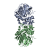
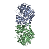
- PDB-4ehb: Crystal structure of the CFTR inhibitory factor Cif with the D129... -
+ Open data

- Basic information
| Entry | Database: PDB / ID: 4ehb | ||||||
|---|---|---|---|---|---|---|---|
| Title | Crystal structure of the CFTR inhibitory factor Cif with the D129S mutation bound to epoxyhexane | ||||||
 Components Components | Putative hydrolase | ||||||
 Keywords Keywords | HYDROLASE / Alpha Beta Hydrolase / Epoxide Hydrolase / secreted | ||||||
| Function / homology |  Function and homology information Function and homology information | ||||||
| Biological species |   Pseudomonas aeruginosa (bacteria) Pseudomonas aeruginosa (bacteria) | ||||||
| Method | X-RAY DIFFRACTION / SYNCHROTRON / PDB ENTRY 3KD2 CHAIN A / Resolution: 1.85 Å | ||||||
 Authors Authors | Hvorecny, K.L. / Bahl, C.D. / Madden, D.R. | ||||||
 Citation Citation | Journal: Biochemistry / Year: 2016 Title: Visualizing the Mechanism of Epoxide Hydrolysis by the Bacterial Virulence Enzyme Cif. Authors: Bahl, C.D. / Hvorecny, K.L. / Morisseau, C. / Gerber, S.A. / Madden, D.R. | ||||||
| History |
|
- Structure visualization
| Structure viewer | Molecule: MolmilJmol/JSmol |
|---|
- Downloads & links
-Download
| PDBx/mmCIF format | 4ehb.cif.gz | 475.2 KB | Display | PDBx/mmCIF format |
|---|---|---|---|---|
| PDB format | pdb4ehb.ent.gz | 406.5 KB | Display | PDB format |
| PDBx/mmJSON format | 4ehb.json.gz | Tree view | PDBx/mmJSON format | |
| Others |  Other downloads Other downloads |
-Validation report
| Arichive directory | https://data.pdbj.org/pub/pdb/validation_reports/eh/4ehbftp://data.pdbj.org/pub/pdb/validation_reports/eh/4ehb | HTTPS FTP |
|---|
-Related structure data


-Links
 PDBj
PDBj

- Assembly
| Deposited unit | 
| ||||||||||||
|---|---|---|---|---|---|---|---|---|---|---|---|---|---|
| 1 | 
| ||||||||||||
| 2 | 
| ||||||||||||
| Unit cell |
| ||||||||||||
| Components on special symmetry positions |
|
-Components
| #1: Protein | Mass: 34136.691 Da / Num. of mol.: 4 / Fragment: Cif / Mutation: D129S Source method: isolated from a genetically manipulated source Source: (gene. exp.) Pseudomonas aeruginosa (bacteria) / Strain: PA14 / Gene: PA14_26090 / Plasmid: pMQ70 / Production host: Escherichia coli (E. coli) / Strain (production host): TOP10 / References: UniProt: Q02P97, UniProt: A0A0H2ZD27*PLUS#2: Chemical | ChemComp-0PZ / (   Mass: 100.159 Da / Num. of mol.: 4 / Source method: obtained synthetically / Formula: C6H12O Mass: 100.159 Da / Num. of mol.: 4 / Source method: obtained synthetically / Formula: C6H12O#3: Water | ChemComp-HOH / | Water Mass: 18.015 Da / Num. of mol.: 1059 / Source method: isolated from a natural source / Formula: H2O Mass: 18.015 Da / Num. of mol.: 1059 / Source method: isolated from a natural source / Formula: H2O |
|---|
-Experimental details
-Experiment
| Experiment | Method: X-RAY DIFFRACTION / Number of used crystals: 1 |
|---|
- Sample preparation
| Crystal | Density Matthews: 2.25 Å3/Da / Density % sol: 45.24 % |
|---|---|
| Crystal grow | Temperature: 291 K / Method: vapor diffusion, hanging drop / pH: 5 Details: 13% PEG 8000, 0.125M calcium chloride, 0.1M sodium acetate, 0.02M epoxyhexane, VAPOR DIFFUSION, HANGING DROP, temperature 291K |
-Data collection
| Diffraction | Mean temperature: 100 K |
|---|---|
| Diffraction source | Source: SYNCHROTRON / Site: NSLS  / Beamline: X6A / Wavelength: 0.9795 Å / Beamline: X6A / Wavelength: 0.9795 Å |
| Detector | Type: ADSC QUANTUM 270 / Detector: CCD / Date: Apr 20, 2011 / Details: Toroidal focusing mirror |
| Radiation | Monochromator: Si(111) channel cut monochromator / Protocol: SINGLE WAVELENGTH / Monochromatic (M) / Laue (L): M / Scattering type: x-ray |
| Radiation wavelength | Wavelength: 0.9795 Å / Relative weight: 1 |
| Reflection | Resolution: 1.85→43.43 Å / Num. obs: 99966 / % possible obs: 96.9 % / Redundancy: 5.8 % / Rsym value: 0.072 / Net I/σ(I): 18.1 |
| Reflection shell | Resolution: 1.85→1.9 Å / Redundancy: 5.8 % / Mean I/σ(I) obs: 4.6 / Num. unique all: 7549 / Rsym value: 0.392 / % possible all: 95.8 |
- Processing
| Software |
| |||||||||||||||||||||||||||||||||||||||||||||||||||||||||||||||||||||||||||||||||||||||||||||||||||||||||||||||||||||||||||||||||||||||||||||||||||||||||||||||||||||||||||||||||||||||||||||||||||||||||||||||||||||||||
|---|---|---|---|---|---|---|---|---|---|---|---|---|---|---|---|---|---|---|---|---|---|---|---|---|---|---|---|---|---|---|---|---|---|---|---|---|---|---|---|---|---|---|---|---|---|---|---|---|---|---|---|---|---|---|---|---|---|---|---|---|---|---|---|---|---|---|---|---|---|---|---|---|---|---|---|---|---|---|---|---|---|---|---|---|---|---|---|---|---|---|---|---|---|---|---|---|---|---|---|---|---|---|---|---|---|---|---|---|---|---|---|---|---|---|---|---|---|---|---|---|---|---|---|---|---|---|---|---|---|---|---|---|---|---|---|---|---|---|---|---|---|---|---|---|---|---|---|---|---|---|---|---|---|---|---|---|---|---|---|---|---|---|---|---|---|---|---|---|---|---|---|---|---|---|---|---|---|---|---|---|---|---|---|---|---|---|---|---|---|---|---|---|---|---|---|---|---|---|---|---|---|---|---|---|---|---|---|---|---|---|---|---|---|---|---|---|---|---|
| Refinement | Method to determine structure: PDB ENTRY 3KD2 CHAIN A / Resolution: 1.85→43.43 Å / SU ML: 0.24 / σ(F): 1.99 / Phase error: 20.82 / Stereochemistry target values: MLHL
| |||||||||||||||||||||||||||||||||||||||||||||||||||||||||||||||||||||||||||||||||||||||||||||||||||||||||||||||||||||||||||||||||||||||||||||||||||||||||||||||||||||||||||||||||||||||||||||||||||||||||||||||||||||||||
| Solvent computation | Shrinkage radii: 0.72 Å / VDW probe radii: 1 Å / Solvent model: FLAT BULK SOLVENT MODEL / Bsol: 38.4 Å2 / ksol: 0.366 e/Å3 | |||||||||||||||||||||||||||||||||||||||||||||||||||||||||||||||||||||||||||||||||||||||||||||||||||||||||||||||||||||||||||||||||||||||||||||||||||||||||||||||||||||||||||||||||||||||||||||||||||||||||||||||||||||||||
| Displacement parameters |
| |||||||||||||||||||||||||||||||||||||||||||||||||||||||||||||||||||||||||||||||||||||||||||||||||||||||||||||||||||||||||||||||||||||||||||||||||||||||||||||||||||||||||||||||||||||||||||||||||||||||||||||||||||||||||
| Refinement step | Cycle: LAST / Resolution: 1.85→43.43 Å
| |||||||||||||||||||||||||||||||||||||||||||||||||||||||||||||||||||||||||||||||||||||||||||||||||||||||||||||||||||||||||||||||||||||||||||||||||||||||||||||||||||||||||||||||||||||||||||||||||||||||||||||||||||||||||
| Refine LS restraints |
| |||||||||||||||||||||||||||||||||||||||||||||||||||||||||||||||||||||||||||||||||||||||||||||||||||||||||||||||||||||||||||||||||||||||||||||||||||||||||||||||||||||||||||||||||||||||||||||||||||||||||||||||||||||||||
| LS refinement shell |
| |||||||||||||||||||||||||||||||||||||||||||||||||||||||||||||||||||||||||||||||||||||||||||||||||||||||||||||||||||||||||||||||||||||||||||||||||||||||||||||||||||||||||||||||||||||||||||||||||||||||||||||||||||||||||
| Refinement TLS params. | Method: refined / Refine-ID: X-RAY DIFFRACTION
| |||||||||||||||||||||||||||||||||||||||||||||||||||||||||||||||||||||||||||||||||||||||||||||||||||||||||||||||||||||||||||||||||||||||||||||||||||||||||||||||||||||||||||||||||||||||||||||||||||||||||||||||||||||||||
| Refinement TLS group |
|
