 Movie
Movie Controller

[English] 日本語
 Yorodumi
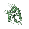
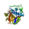
- PDB-4ds1: The Structure of a Yeast Dyn2-Nup159 Complex and the Molecular Ba... -
+ Open data

- Basic information
| Entry | Database: PDB / ID: 4ds1 | ||||||
|---|---|---|---|---|---|---|---|
| Title | The Structure of a Yeast Dyn2-Nup159 Complex and the Molecular Basis for the Dynein Light Chain - Nuclear Pore Interaction | ||||||
 Components Components |
| ||||||
 Keywords Keywords |  STRUCTURAL PROTEIN/TRANSPORT PROTEIN / nucleoporin / Dynein Light Chain fold / Peptide Binding / Nuclear Pore / STRUCTURAL PROTEIN-TRANSPORT PROTEIN complex STRUCTURAL PROTEIN/TRANSPORT PROTEIN / nucleoporin / Dynein Light Chain fold / Peptide Binding / Nuclear Pore / STRUCTURAL PROTEIN-TRANSPORT PROTEIN complex | ||||||
| Function / homology |  Function and homology information Function and homology information: / peroxisomal importomer complex / adenyl-nucleotide exchange factor activity / nuclear pore localization / nuclear pore central transport channel / establishment of mitotic spindle localization / nuclear migration along microtubule / nuclear pore complex assembly / nuclear pore cytoplasmic filaments / cytoplasmic dynein complex ...: / peroxisomal importomer complex / adenyl-nucleotide exchange factor activity / nuclear pore localization / nuclear pore central transport channel / establishment of mitotic spindle localization / nuclear migration along microtubule / nuclear pore complex assembly / nuclear pore cytoplasmic filaments / cytoplasmic dynein complex / structural constituent of nuclear pore / poly(A)+ mRNA export from nucleus / nucleocytoplasmic transport / NLS-bearing protein import into nucleus / dynein intermediate chain binding / establishment of mitotic spindle orientation / ribosomal small subunit export from nucleus / ribosomal large subunit export from nucleus / mRNA transport / cytoplasmic microtubule / nuclear pore / Neutrophil degranulation / protein export from nucleus / nuclear periphery / transcription corepressor activity / protein transport / nuclear envelope / nuclear membrane / molecular adaptor activity / protein-containing complex binding / nucleus / cytoplasmSimilarity search - Function | ||||||
| Biological species |  Saccharomyces cerevisiae (brewer's yeast) Saccharomyces cerevisiae (brewer's yeast) | ||||||
| Method | X-RAY DIFFRACTION / MOLECULAR REPLACEMENT / Resolution: 1.851 Å | ||||||
 Authors Authors | Slep, K.C. / Romes, E.M. | ||||||
 Citation Citation | Journal: J.Biol.Chem. / Year: 2012 Title: Structure of a yeast Dyn2-Nup159 complex and molecular basis for dynein light chain-nuclear pore interaction. Authors: Romes, E.M. / Tripathy, A. / Slep, K.C. | ||||||
| History |
|
- Structure visualization
| Structure viewer | Molecule: MolmilJmol/JSmol |
|---|
- Downloads & links
-Download
| PDBx/mmCIF format | 4ds1.cif.gz | 55.7 KB | Display | PDBx/mmCIF format |
|---|---|---|---|---|
| PDB format | pdb4ds1.ent.gz | 39.8 KB | Display | PDB format |
| PDBx/mmJSON format | 4ds1.json.gz | Tree view | PDBx/mmJSON format | |
| Others |  Other downloads Other downloads |
-Validation report
| Arichive directory | https://data.pdbj.org/pub/pdb/validation_reports/ds/4ds1ftp://data.pdbj.org/pub/pdb/validation_reports/ds/4ds1 | HTTPS FTP |
|---|
-Related structure data
| Related structure data | 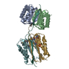 2pg1S S: Starting model for refinement |
|---|---|
| Similar structure data |


-Links
 PDBj
PDBj


- Assembly
| Deposited unit | 
| ||||||||
|---|---|---|---|---|---|---|---|---|---|
| 1 |
| ||||||||
| Unit cell |
|
-Components
| #1: Protein | Mass: 10868.436 Da / Num. of mol.: 2 Source method: isolated from a genetically manipulated source Source: (gene. exp.) Saccharomyces cerevisiae (brewer's yeast)Strain: S288C / Gene: DYN2, SLC1, YDR424C / Plasmid: pGEX-6P2 / Production host:  Escherichia coli (E. coli) / Strain (production host): BL21 DE3 pLysS / References: UniProt: Q02647 Escherichia coli (E. coli) / Strain (production host): BL21 DE3 pLysS / References: UniProt: Q02647#2: Protein/peptide | Mass: 1210.249 Da / Num. of mol.: 2 / Fragment: UNP residues 1116-1126 / Source method: obtained synthetically / Source: (synth.) Saccharomyces cerevisiae (brewer's yeast) / References: UniProt: P40477#3: Water | ChemComp-HOH / | Water Mass: 18.015 Da / Num. of mol.: 217 / Source method: isolated from a natural source / Formula: H2O Mass: 18.015 Da / Num. of mol.: 217 / Source method: isolated from a natural source / Formula: H2O |
|---|
-Experimental details
-Experiment
| Experiment | Method: X-RAY DIFFRACTION / Number of used crystals: 1 |
|---|
- Sample preparation
| Crystal | Density Matthews: 1.86 Å3/Da / Density % sol: 33.92 % |
|---|---|
| Crystal grow | Temperature: 293 K / Method: vapor diffusion, hanging drop / pH: 5.5 Details: 0.3 M Ammonium acetate, 5% methyl pentanediol (v/v), 35% PEG 4000 (w/v), pH 5.5, VAPOR DIFFUSION, HANGING DROP, temperature 293K |
-Data collection
| Diffraction | Mean temperature: 100 K |
|---|---|
| Diffraction source | Source: ROTATING ANODE / Type: RIGAKU MICROMAX-007 HF / Wavelength: 1.54 Å |
| Detector | Type: RIGAKU SATURN 944+ / Detector: CCD / Date: Jan 25, 2010 / Details: Osmic Mirrors |
| Radiation | Monochromator: copper Kalpha / Osmic mirrors / Protocol: SINGLE WAVELENGTH / Monochromatic (M) / Laue (L): M / Scattering type: x-ray |
| Radiation wavelength | Wavelength: 1.54 Å / Relative weight: 1 |
| Reflection | Resolution: 1.85→29.2 Å / Num. obs: 15081 / % possible obs: 93.9 % / Observed criterion σ(F): 0 / Observed criterion σ(I): -3 / Redundancy: 5.4 % / Biso Wilson estimate: 15.71 Å2 / Rmerge(I) obs: 0.045 / Rsym value: 0.045 / Net I/σ(I): 24.8 |
| Reflection shell | Resolution: 1.85→1.92 Å / Redundancy: 2.5 % / Mean I/σ(I) obs: 5.9 / Rsym value: 0.15 / % possible all: 62.7 |
- Processing
| Software |
| |||||||||||||||||||||||||||||||||||||||||||||||||||||||||||||||||||||||||||||
|---|---|---|---|---|---|---|---|---|---|---|---|---|---|---|---|---|---|---|---|---|---|---|---|---|---|---|---|---|---|---|---|---|---|---|---|---|---|---|---|---|---|---|---|---|---|---|---|---|---|---|---|---|---|---|---|---|---|---|---|---|---|---|---|---|---|---|---|---|---|---|---|---|---|---|---|---|---|---|
| Refinement | Method to determine structure: MOLECULAR REPLACEMENT Starting model: PDB entry 2PG1 lacking bound peptide Resolution: 1.851→29.249 Å / SU ML: 0.17 / σ(F): 0 / Phase error: 16.68 / Stereochemistry target values: ML
| |||||||||||||||||||||||||||||||||||||||||||||||||||||||||||||||||||||||||||||
| Solvent computation | Shrinkage radii: 0.73 Å / VDW probe radii: 1 Å / Solvent model: FLAT BULK SOLVENT MODEL / Bsol: 36.9 Å2 / ksol: 0.36 e/Å3 | |||||||||||||||||||||||||||||||||||||||||||||||||||||||||||||||||||||||||||||
| Displacement parameters |
| |||||||||||||||||||||||||||||||||||||||||||||||||||||||||||||||||||||||||||||
| Refinement step | Cycle: LAST / Resolution: 1.851→29.249 Å
| |||||||||||||||||||||||||||||||||||||||||||||||||||||||||||||||||||||||||||||
| Refine LS restraints |
| |||||||||||||||||||||||||||||||||||||||||||||||||||||||||||||||||||||||||||||
| LS refinement shell |
|
