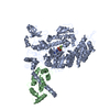
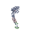
regulation of slow-twitch skeletal muscle fiber contraction / regulation of the force of skeletal muscle contraction / muscle myosin complex / muscle filament sliding / regulation of the force of heart contraction / transition between fast and slow fiber / myosin filament / myosin II complex / adult heart development / cardiac muscle hypertrophy in response to stress ...regulation of slow-twitch skeletal muscle fiber contraction / regulation of the force of skeletal muscle contraction / muscle myosin complex / muscle filament sliding / regulation of the force of heart contraction / transition between fast and slow fiber / myosin filament / myosin II complex / adult heart development / cardiac muscle hypertrophy in response to stress / myosin complex / sarcomere organization / microfilament motor activity / ventricular cardiac muscle tissue morphogenesis / myofibril / skeletal muscle contraction / striated muscle contraction / ATP metabolic process / stress fiber / cardiac muscle contraction / regulation of heart rate / sarcomere / muscle contraction / Z disc / actin filament binding / calmodulin binding / ATP binding / cytoplasm Similarity search - Function
DNA repair protein XRCC4-like, C-terminal / Myosin tail / Myosin tail / Myosin N-terminal SH3-like domain / Myosin S1 fragment, N-terminal / Myosin, N-terminal, SH3-like / Myosin N-terminal SH3-like domain profile. / Short calmodulin-binding motif containing conserved Ile and Gln residues. / Myosin head, motor domain / Myosin head (motor domain) ...DNA repair protein XRCC4-like, C-terminal / Myosin tail / Myosin tail / Myosin N-terminal SH3-like domain / Myosin S1 fragment, N-terminal / Myosin, N-terminal, SH3-like / Myosin N-terminal SH3-like domain profile. / Short calmodulin-binding motif containing conserved Ile and Gln residues. / Myosin head, motor domain / Myosin head (motor domain) / Myosin motor domain profile. / Myosin. Large ATPases. / IQ motif profile. / IQ motif, EF-hand binding site / Kinesin motor domain superfamily / P-loop containing nucleoside triphosphate hydrolase Similarity search - Domain/homology
Resolution: 2.6→25 Å / Cor.coef. Fo:Fc: 0.935 / Cor.coef. Fo:Fc free: 0.896 / WRfactor Rfree: 0.2364 / WRfactor Rwork: 0.1924 / Occupancy max: 1 / Occupancy min: 0.3 / FOM work R set: 0.8105 / SU B: 20.348 / SU ML: 0.219 / SU R Cruickshank DPI: 0.5907 / SU Rfree: 0.3086 / Cross valid method: THROUGHOUT / σ(F): 0 / ESU R: 0.591 / ESU R Free: 0.309 / Stereochemistry target values: MAXIMUM LIKELIHOOD Details: HYDROGENS HAVE BEEN ADDED IN THE RIDING POSITIONS. ATOM RECORD CONTAINS SUM OF TLS AND RESIDUAL B FACTORS. ANISOU RECORD CONTAINS SUM OF TLS AND RESIDUAL U FACTORS.
Rfactor
Num. reflection
% reflection
Selection details
Rfree
0.2572
2959
5.1 %
RANDOM
Rwork
0.21
-
-
-
all
0.2124
55769
-
-
obs
0.2124
55440
99.41 %
-
Solvent computation
Ion probe radii: 0.8 Å / Shrinkage radii: 0.8 Å / VDW probe radii: 1.1 Å / Solvent model: MASK
In the structure databanks used in Yorodumi, some data are registered as the other names, "COVID-19 virus" and "2019-nCoV". Here are the details of the virus and the list of structure data.
Jan 31, 2019. EMDB accession codes are about to change! (news from PDBe EMDB page)
EMDB accession codes are about to change! (news from PDBe EMDB page)
The allocation of 4 digits for EMDB accession codes will soon come to an end. Whilst these codes will remain in use, new EMDB accession codes will include an additional digit and will expand incrementally as the available range of codes is exhausted. The current 4-digit format prefixed with “EMD-” (i.e. EMD-XXXX) will advance to a 5-digit format (i.e. EMD-XXXXX), and so on. It is currently estimated that the 4-digit codes will be depleted around Spring 2019, at which point the 5-digit format will come into force.
The EM Navigator/Yorodumi systems omit the EMD- prefix.
Related info.:Q: What is EMD? / ID/Accession-code notation in Yorodumi/EM Navigator
Yorodumi is a browser for structure data from EMDB, PDB, SASBDB, etc.
This page is also the successor to EM Navigator detail page, and also detail information page/front-end page for Omokage search.
The word "yorodu" (or yorozu) is an old Japanese word meaning "ten thousand". "mi" (miru) is to see.
Related info.:EMDB / PDB / SASBDB / Comparison of 3 databanks / Yorodumi Search / Aug 31, 2016. New EM Navigator & Yorodumi / Yorodumi Papers / Jmol/JSmol / Function and homology information / Changes in new EM Navigator and Yorodumi
 Movie
Movie Controller


 Open data
 Basic information
 Components
 Keywords
 Function and homology information
 Authors
 Citation
 Structure visualization
 Downloads & links
 Other downloads


 PDBj
PDBj


 Assembly


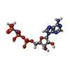
 Mass: 506.196 Da / Num. of mol.: 2 / Source method: obtained synthetically / Formula: C10H17N6O12P3 / Comment: AMP-PNP, energy-carrying molecule analogue*YM
 Mass: 54.938 Da / Num. of mol.: 2 / Source method: obtained synthetically / Formula: Mn
 Mass: 18.015 Da / Num. of mol.: 202 / Source method: isolated from a natural source / Formula: H2O
 Sample preparation
 / Beamline: 19-ID / Wavelength: 0.97907 Å
 Processing