 Movie
Movie Controller

[English] 日本語
 Yorodumi
- PDB-4bvb: CRYSTAL STRUCTURE OF HUMAN SIRT3 IN COMPLEX WITH THE INHIBITOR EX... -
+ Open data

- Basic information
| Entry | Database: PDB / ID: 4bvb | ||||||
|---|---|---|---|---|---|---|---|
| Title | CRYSTAL STRUCTURE OF HUMAN SIRT3 IN COMPLEX WITH THE INHIBITOR EX-527 AND ADP-RIBOSE | ||||||
 Components Components | NAD-DEPENDENT PROTEIN DEACETYLASE SIRTUIN-3, MITOCHONDRIAL | ||||||
 Keywords Keywords |  HYDROLASE / NAD-DEPENDENT DEACETYLASE / INHIBITOR COMPLEX / ADP-RIBOSE HYDROLASE / NAD-DEPENDENT DEACETYLASE / INHIBITOR COMPLEX / ADP-RIBOSE | ||||||
| Function / homology |  Function and homology information Function and homology informationpositive regulation of catalase activity / positive regulation of ceramide biosynthetic process / peptidyl-lysine deacetylation / positive regulation of superoxide dismutase activity / NAD-dependent protein lysine deacetylase activity / protein acetyllysine N-acetyltransferase / NAD-dependent histone deacetylase activity / protein deacetylation / Regulation of FOXO transcriptional activity by acetylation / NAD+ binding ...positive regulation of catalase activity / positive regulation of ceramide biosynthetic process / peptidyl-lysine deacetylation / positive regulation of superoxide dismutase activity / NAD-dependent protein lysine deacetylase activity / protein acetyllysine N-acetyltransferase / NAD-dependent histone deacetylase activity / protein deacetylation / Regulation of FOXO transcriptional activity by acetylation / NAD+ binding / negative regulation of reactive oxygen species metabolic process / FOXO-mediated transcription of oxidative stress, metabolic and neuronal genes / aerobic respiration / Transcriptional activation of mitochondrial biogenesis / negative regulation of ERK1 and ERK2 cascade / positive regulation of insulin secretion / transferase activity / sequence-specific DNA binding / mitochondrial matrix / enzyme binding / protein-containing complex / mitochondrion / zinc ion binding / nucleoplasm / nucleusSimilarity search - Function | ||||||
| Biological species |  HOMO SAPIENS (human) HOMO SAPIENS (human) | ||||||
| Method | X-RAY DIFFRACTION / SYNCHROTRON / MOLECULAR REPLACEMENT / Resolution: 2 Å | ||||||
 Authors Authors | Gertz, M. / Weyand, M. / Steegborn, C. | ||||||
 Citation Citation | Journal: Proc.Natl.Acad.Sci.USA / Year: 2013 Title: Ex-527 Inhibits Sirtuins by Exploiting Their Unique Nad+-Dependent Deacetylation Mechanism Authors: Gertz, M. / Fischer, F. / Nguyen, G.T.T. / Lakshminarasimhan, M. / Schutkowski, M. / Weyand, M. / Steegborn, C. | ||||||
| History |
|
- Structure visualization
| Structure viewer | Molecule: MolmilJmol/JSmol |
|---|
- Downloads & links
-Download
| PDBx/mmCIF format | 4bvb.cif.gz | 76.2 KB | Display | PDBx/mmCIF format |
|---|---|---|---|---|
| PDB format | pdb4bvb.ent.gz | 53.9 KB | Display | PDB format |
| PDBx/mmJSON format | 4bvb.json.gz | Tree view | PDBx/mmJSON format | |
| Others |  Other downloads Other downloads |
-Validation report
| Arichive directory | https://data.pdbj.org/pub/pdb/validation_reports/bv/4bvbftp://data.pdbj.org/pub/pdb/validation_reports/bv/4bvb | HTTPS FTP |
|---|
-Related structure data
| Related structure data |  4buzC  4bv2C  4bv3SC  4bveC  4bvfC  4bvgC 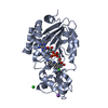 4bvhC C: citing same article ( S: Starting model for refinement |
|---|---|
| Similar structure data |


-Links
 PDBj
PDBj


- Assembly
| Deposited unit | 
| ||||||||
|---|---|---|---|---|---|---|---|---|---|
| 1 |
| ||||||||
| Unit cell |
|
-Components
| #1: Protein | Mass: 31484.219 Da / Num. of mol.: 1 Source method: isolated from a genetically manipulated source Source: (gene. exp.) HOMO SAPIENS (human) / Plasmid: PVFT3S / Production host:  ESCHERICHIA COLI (E. coli) / Strain (production host): BL21(DE3) / Variant (production host): ROSETTA2 ESCHERICHIA COLI (E. coli) / Strain (production host): BL21(DE3) / Variant (production host): ROSETTA2References: UniProt: Q9NTG7, Hydrolases; Acting on carbon-nitrogen bonds, other than peptide bonds; In linear amides |
|---|---|
| #2: Chemical | ChemComp-OCZ / (  Mass: 248.708 Da / Num. of mol.: 1 / Source method: obtained synthetically / Formula: C13H13ClN2O Mass: 248.708 Da / Num. of mol.: 1 / Source method: obtained synthetically / Formula: C13H13ClN2O |
| #3: Chemical | ChemComp-AR6 / [(  Mass: 559.316 Da / Num. of mol.: 1 / Source method: obtained synthetically / Formula: C15H23N5O14P2 Mass: 559.316 Da / Num. of mol.: 1 / Source method: obtained synthetically / Formula: C15H23N5O14P2 |
| #4: Chemical | ChemComp-ZN /   Mass: 65.409 Da / Num. of mol.: 1 / Source method: obtained synthetically / Formula: Zn Mass: 65.409 Da / Num. of mol.: 1 / Source method: obtained synthetically / Formula: Zn |
| #5: Water | ChemComp-HOH / Water Mass: 18.015 Da / Num. of mol.: 149 / Source method: isolated from a natural source / Formula: H2O Mass: 18.015 Da / Num. of mol.: 149 / Source method: isolated from a natural source / Formula: H2O |
| Sequence details | CONSTRUCT COVERS RESIDUES 116-399 |
-Experimental details
-Experiment
| Experiment | Method: X-RAY DIFFRACTION / Number of used crystals: 1 |
|---|
- Sample preparation
| Crystal | Density Matthews: 2.12 Å3/Da / Density % sol: 42.11 % / Description: NONE |
|---|---|
| Crystal grow | Details: 0.08 M KH2PO4 18 % PEG 8000 20% GLYCEROL |
-Data collection
| Diffraction | Mean temperature: 100 K |
|---|---|
| Diffraction source | Source: SYNCHROTRON / Site: BESSY  / Beamline: 14.1 / Wavelength: 0.918 / Beamline: 14.1 / Wavelength: 0.918 |
| Detector | Type: MARMOSAIC 225 mm CCD / Detector: CCD / Date: Nov 29, 2011 / Details: COLLIMATOR |
| Radiation | Monochromator: SI(111) MONOCHROMATOR / Protocol: SINGLE WAVELENGTH / Monochromatic (M) / Laue (L): M / Scattering type: x-ray |
| Radiation wavelength | Wavelength: 0.918 Å / Relative weight: 1 |
| Reflection | Resolution: 2→46.6 Å / Num. obs: 18341 / % possible obs: 98.1 % / Observed criterion σ(I): -3 / Redundancy: 4 % / Rmerge(I) obs: 0.07 / Net I/σ(I): 18.6 |
| Reflection shell | Resolution: 2→2.05 Å / Redundancy: 3.5 % / Rmerge(I) obs: 0.37 / Mean I/σ(I) obs: 3.9 / % possible all: 93.1 |
- Processing
| Software |
| ||||||||||||||||||||||||||||||||||||||||||||||||||||||||||||||||||||||||||||||||||||||||||||||||||||||||||||||||||||||||||||||||||||||||||||||||||||||||||||||||||||||||||||||||||||||
|---|---|---|---|---|---|---|---|---|---|---|---|---|---|---|---|---|---|---|---|---|---|---|---|---|---|---|---|---|---|---|---|---|---|---|---|---|---|---|---|---|---|---|---|---|---|---|---|---|---|---|---|---|---|---|---|---|---|---|---|---|---|---|---|---|---|---|---|---|---|---|---|---|---|---|---|---|---|---|---|---|---|---|---|---|---|---|---|---|---|---|---|---|---|---|---|---|---|---|---|---|---|---|---|---|---|---|---|---|---|---|---|---|---|---|---|---|---|---|---|---|---|---|---|---|---|---|---|---|---|---|---|---|---|---|---|---|---|---|---|---|---|---|---|---|---|---|---|---|---|---|---|---|---|---|---|---|---|---|---|---|---|---|---|---|---|---|---|---|---|---|---|---|---|---|---|---|---|---|---|---|---|---|---|
| Refinement | Method to determine structure: MOLECULAR REPLACEMENT Starting model: PDB ENTRY 4BV3 Resolution: 2→46.61 Å / Cor.coef. Fo:Fc: 0.954 / Cor.coef. Fo:Fc free: 0.92 / SU B: 4.062 / SU ML: 0.114 / Cross valid method: THROUGHOUT / ESU R: 0.194 / ESU R Free: 0.176 / Stereochemistry target values: MAXIMUM LIKELIHOOD Details: HYDROGENS HAVE BEEN ADDED IN THE RIDING POSITIONS. HYDROGENS HAVE BEEN USED IF PRESENT IN THE INPUT. U VALUES REFINED INDIVIDUALLY
| ||||||||||||||||||||||||||||||||||||||||||||||||||||||||||||||||||||||||||||||||||||||||||||||||||||||||||||||||||||||||||||||||||||||||||||||||||||||||||||||||||||||||||||||||||||||
| Solvent computation | Ion probe radii: 0.8 Å / Shrinkage radii: 0.8 Å / VDW probe radii: 1.2 Å / Solvent model: MASK | ||||||||||||||||||||||||||||||||||||||||||||||||||||||||||||||||||||||||||||||||||||||||||||||||||||||||||||||||||||||||||||||||||||||||||||||||||||||||||||||||||||||||||||||||||||||
| Displacement parameters | Biso mean: 19.548 Å2
| ||||||||||||||||||||||||||||||||||||||||||||||||||||||||||||||||||||||||||||||||||||||||||||||||||||||||||||||||||||||||||||||||||||||||||||||||||||||||||||||||||||||||||||||||||||||
| Refinement step | Cycle: LAST / Resolution: 2→46.61 Å
| ||||||||||||||||||||||||||||||||||||||||||||||||||||||||||||||||||||||||||||||||||||||||||||||||||||||||||||||||||||||||||||||||||||||||||||||||||||||||||||||||||||||||||||||||||||||
| Refine LS restraints |
|