- PDB-4a15: Crystal structure of an XPD DNA complex -
+
Open data
ID or keywords:
Loading...
-
Basic information
Entry
Database: PDB / ID: 4a15
Title
Crystal structure of an XPD DNA complex
Components
5'-D(*DTP*AP*CP*GP)-3'
ATP-DEPENDENT DNA HELICASE TA0057
Keywords
HYDROLASE / HELICASE / NUCLEOTIDE EXCISION REPAIR
Function / homology
Function and homology information
DNA helicase activity / 4 iron, 4 sulfur cluster binding / DNA helicase / DNA repair / ATP hydrolysis activity / DNA binding / ATP binding / metal ion binding Similarity search - Function
Resolution: 2.2→39.53 Å / Cor.coef. Fo:Fc: 0.952 / Cor.coef. Fo:Fc free: 0.929 / SU B: 13.859 / SU ML: 0.179 / Cross valid method: THROUGHOUT / ESU R: 0.318 / ESU R Free: 0.233 / Stereochemistry target values: MAXIMUM LIKELIHOOD Details: HYDROGENS HAVE BEEN ADDED IN THE RIDING POSITIONS. U VALUES WITH TLS ADDED.
Rfactor
Num. reflection
% reflection
Selection details
Rfree
0.25049
1545
5 %
RANDOM
Rwork
0.1923
-
-
-
obs
0.19524
29635
99.02 %
-
Solvent computation
Ion probe radii: 0.8 Å / Shrinkage radii: 0.8 Å / VDW probe radii: 1.2 Å / Solvent model: BABINET MODEL WITH MASK
 Movie
Movie Controller


 Open data
 Basic information
 Components
 Keywords
 HYDROLASE /
  Function and homology information

 Authors
 Citation
 Structure visualization
 Downloads & links
 Other downloads
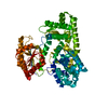


 PDBj
PDBj Assembly


 Mass: 351.640 Da / Num. of mol.: 1 / Source method: obtained synthetically / Formula: Fe4S4
 Mass: 96.063 Da / Num. of mol.: 3 / Source method: obtained synthetically / Formula: SO4
 Mass: 40.078 Da / Num. of mol.: 1 / Source method: obtained synthetically / Formula: Ca
 Sample preparation
 / Beamline: BM14 / Wavelength: 0.972
  Processing