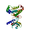
[English] 日本語
Yorodumi
- PDB-3zfy: Crystal structure of EphB3 -

+
Open data


ID or keywords:

Loading...

-
Basic information

Entry
Database: PDB / ID: 3zfy
TitleCrystal structure of EphB3
ComponentsEPHRIN TYPE-B RECEPTOR 3
KeywordsTRANSFERASE
Function / homology
Function and homology information


urogenital system development / ephrin receptor activity / axon guidance receptor activity / central nervous system projection neuron axonogenesis / regulation of cell-cell adhesion / dendritic spine development / corpus callosum development / transmembrane-ephrin receptor activity / dendritic spine morphogenesis / retinal ganglion cell axon guidance ...urogenital system development / ephrin receptor activity / axon guidance receptor activity / central nervous system projection neuron axonogenesis / regulation of cell-cell adhesion / dendritic spine development / corpus callosum development / transmembrane-ephrin receptor activity / dendritic spine morphogenesis / retinal ganglion cell axon guidance / axonal fasciculation / positive regulation of synapse assembly / EPH-Ephrin signaling / Ephrin signaling / digestive tract morphogenesis / regulation of axonogenesis / regulation of GTPase activity / roof of mouth development / EPH-ephrin mediated repulsion of cells / ephrin receptor signaling pathway / EPHB-mediated forward signaling / substrate adhesion-dependent cell spreading / thymus development / axon guidance / receptor protein-tyrosine kinase / cell migration / angiogenesis / protein autophosphorylation / dendrite / extracellular region / ATP binding / plasma membrane / cytosol
Similarity search - Function
Ephrin type-B receptor 3, ligand binding domain / Tyrosine-protein kinase ephrin type A/B receptor-like / Tyrosine-protein kinase ephrin type A/B receptor-like / Ephrin receptor type-A /type-B / Ephrin receptor ligand binding domain / Tyrosine-protein kinase, receptor class V, conserved site / Ephrin receptor, transmembrane domain / Ephrin receptor ligand binding domain / Ephrin type-A receptor 2 transmembrane domain / Receptor tyrosine kinase class V signature 1. ...Ephrin type-B receptor 3, ligand binding domain / Tyrosine-protein kinase ephrin type A/B receptor-like / Tyrosine-protein kinase ephrin type A/B receptor-like / Ephrin receptor type-A /type-B / Ephrin receptor ligand binding domain / Tyrosine-protein kinase, receptor class V, conserved site / Ephrin receptor, transmembrane domain / Ephrin receptor ligand binding domain / Ephrin type-A receptor 2 transmembrane domain / Receptor tyrosine kinase class V signature 1. / Receptor tyrosine kinase class V signature 2. / Eph receptor ligand-binding domain profile. / Ephrin receptor ligand binding domain / Putative ephrin-receptor like / SAM domain (Sterile alpha motif) / SAM domain profile. / Sterile alpha motif. / Sterile alpha motif domain / Sterile alpha motif/pointed domain superfamily / Fibronectin type III domain / Fibronectin type 3 domain / Fibronectin type-III domain profile. / Galactose-binding-like domain superfamily / Fibronectin type III / Fibronectin type III superfamily / Tyrosine-protein kinase, catalytic domain / Tyrosine kinase, catalytic domain / Tyrosine protein kinases specific active-site signature. / Tyrosine-protein kinase, active site / Protein tyrosine and serine/threonine kinase / Serine-threonine/tyrosine-protein kinase, catalytic domain / Transferase(Phosphotransferase) domain 1 / Transferase(Phosphotransferase); domain 1 / Phosphorylase Kinase; domain 1 / Phosphorylase Kinase; domain 1 / Protein kinase, ATP binding site / Protein kinases ATP-binding region signature. / Immunoglobulin-like fold / Protein kinase domain profile. / Protein kinase domain / Protein kinase-like domain superfamily / 2-Layer Sandwich / Orthogonal Bundle / Mainly Alpha / Alpha Beta
Similarity search - Domain/homology
Ephrin type-B receptor 3
Similarity search - Component
Biological speciesHOMO SAPIENS (human)
MethodX-RAY DIFFRACTION / SYNCHROTRON / MOLECULAR REPLACEMENT / Resolution: 2.2 Å
AuthorsDebreczeni, J.E. / Overman, R. / Truman, C. / McAlister, M. / Attwood, T.K.
CitationJournal: Protein Sci. / Year: 2014
Title: Completing the Structural Family Portrait of the Human Ephb Tyrosine Kinase Domains
Authors: Overman, R.C. / Debreczeni, J.E. / Truman, C.M. / Mcalister, M.S. / Attwood, T.K.
History
DepositionDec 12, 2012Deposition site: PDBE / Processing site: PDBE
Revision 1.0Jan 8, 2014Provider: repository / Type: Initial release
Revision 1.1Mar 5, 2014Group: Database references
Revision 1.2Apr 9, 2014Group: Database references
Revision 1.3May 7, 2014Group: Database references
Revision 1.4May 8, 2024Group: Data collection / Database references / Other
Category: chem_comp_atom / chem_comp_bond ...chem_comp_atom / chem_comp_bond / database_2 / pdbx_database_status
Item: _database_2.pdbx_DOI / _database_2.pdbx_database_accession / _pdbx_database_status.status_code_sf

-
Structure visualization

Structure viewerMolecule:
MolmilJmol/JSmol

Downloads & links

-
Assembly

Deposited unit
A: EPHRIN TYPE-B RECEPTOR 3
B: EPHRIN TYPE-B RECEPTOR 3


Theoretical massNumber of molelcules
Total (without water)67,5312
Polymers67,5312
Non-polymers00
Water2,306128
1
A: EPHRIN TYPE-B RECEPTOR 3


Theoretical massNumber of molelcules
Total (without water)33,7661
Polymers33,7661
Non-polymers00
Water181
TypeNameSymmetry operationNumber
identity operation1_555x,y,z1
MethodPISA
2
B: EPHRIN TYPE-B RECEPTOR 3


Theoretical massNumber of molelcules
Total (without water)33,7661
Polymers33,7661
Non-polymers00
Water181
TypeNameSymmetry operationNumber
identity operation1_555x,y,z1
MethodPISA
Unit cell
Length a, b, c (Å)46.990, 56.488, 61.354
Angle α, β, γ (deg.)93.02, 90.65, 90.03
Int Tables number1
Space group name H-MP1

-
Components

#1: Protein EPHRIN TYPE-B RECEPTOR 3 / EPH-LIKE TYROSINE KINASE 2 / EPH-LIKE KINASE 2 / EMBRYONIC KINASE 2 / EK2 / HEK2 / TYROSINE-PROTEIN ...EPH-LIKE TYROSINE KINASE 2 / EPH-LIKE KINASE 2 / EMBRYONIC KINASE 2 / EK2 / HEK2 / TYROSINE-PROTEIN KINASE TYRO6 / EPHB3


Mass: 33765.711 Da / Num. of mol.: 2 / Fragment: KINASE DOMAIN, RESIDUES 616-910
Source method: isolated from a genetically manipulated source
Source: (gene. exp.) HOMO SAPIENS (human) / Production host: ESCHERICHIA COLI (E. coli) / Strain (production host): BL21(DE3) / Variant (production host): STAR
References: UniProt: P54753, receptor protein-tyrosine kinase
#2: Water ChemComp-HOH / water / Water


Mass: 18.015 Da / Num. of mol.: 128 / Source method: isolated from a natural source / Formula: H2O

-
Experimental details

-
Experiment

ExperimentMethod: X-RAY DIFFRACTION / Number of used crystals: 1

-
Sample preparation

CrystalDensity Matthews: 2.32 Å3/Da / Density % sol: 47 % / Description: NONE

-
Data collection

DiffractionMean temperature: 100 K
Diffraction sourceSource: SYNCHROTRON / Site: Diamond / Beamline: I04-1 / Wavelength: 0.979
DetectorType: ADSC QUANTUM 315 / Detector: CCD / Date: Jul 13, 2011
RadiationProtocol: SINGLE WAVELENGTH / Monochromatic (M) / Laue (L): M / Scattering type: x-ray
Radiation wavelengthWavelength: 0.979 Å / Relative weight: 1
ReflectionResolution: 2.2→61.2 Å / Num. obs: 29560 / % possible obs: 92.1 % / Observed criterion σ(I): 2 / Redundancy: 2 % / Rmerge(I) obs: 0.05 / Net I/σ(I): 9.68
Reflection shellResolution: 2.2→2.28 Å / Redundancy: 1.9 % / Rmerge(I) obs: 0.14 / Mean I/σ(I) obs: 3.83 / % possible all: 83

-
Processing

Software
NameVersionClassification
REFMAC5.6.0117refinement
XDSdata reduction
SCALAdata scaling
PHASERphasing
RefinementMethod to determine structure: MOLECULAR REPLACEMENT / Resolution: 2.2→61.26 Å / Cor.coef. Fo:Fc: 0.92 / Cor.coef. Fo:Fc free: 0.901 / SU B: 13.171 / SU ML: 0.169 / Cross valid method: THROUGHOUT / ESU R: 0.307 / ESU R Free: 0.222 / Stereochemistry target values: MAXIMUM LIKELIHOOD
Details: HYDROGENS HAVE BEEN ADDED IN THE RIDING POSITIONS. U VALUES WITH TLS ADDED
RfactorNum. reflection% reflectionSelection details
Rfree0.25188 1515 5.1 %RANDOM
Rwork0.22406 ---
obs0.22552 28046 92.3 %-
Solvent computationIon probe radii: 0.8 Å / Shrinkage radii: 0.8 Å / VDW probe radii: 1.2 Å / Solvent model: MASK
Displacement parametersBiso mean: 33.017 Å2
Baniso -1Baniso -2Baniso -3
1--2.78 Å20.18 Å20.09 Å2
2---1.99 Å2-0.18 Å2
3---4.75 Å2
Refinement stepCycle: LAST / Resolution: 2.2→61.26 Å
ProteinNucleic acidLigandSolventTotal
Num. atoms3970 0 0 128 4098
Refine LS restraints
Refine-IDTypeDev idealDev ideal targetNumber
X-RAY DIFFRACTIONr_bond_refined_d0.0090.0194070
X-RAY DIFFRACTIONr_bond_other_d0.0020.022701
X-RAY DIFFRACTIONr_angle_refined_deg1.2741.9595532
X-RAY DIFFRACTIONr_angle_other_deg0.93436571
X-RAY DIFFRACTIONr_dihedral_angle_1_deg5.8945508
X-RAY DIFFRACTIONr_dihedral_angle_2_deg34.62323.736174
X-RAY DIFFRACTIONr_dihedral_angle_3_deg14.35315658
X-RAY DIFFRACTIONr_dihedral_angle_4_deg18.1671528
X-RAY DIFFRACTIONr_chiral_restr0.0680.2636
X-RAY DIFFRACTIONr_gen_planes_refined0.0050.0214500
X-RAY DIFFRACTIONr_gen_planes_other0.0020.02832
X-RAY DIFFRACTIONr_nbd_refined
X-RAY DIFFRACTIONr_nbd_other
X-RAY DIFFRACTIONr_nbtor_refined
X-RAY DIFFRACTIONr_nbtor_other
X-RAY DIFFRACTIONr_xyhbond_nbd_refined
X-RAY DIFFRACTIONr_xyhbond_nbd_other
X-RAY DIFFRACTIONr_metal_ion_refined
X-RAY DIFFRACTIONr_metal_ion_other
X-RAY DIFFRACTIONr_symmetry_vdw_refined
X-RAY DIFFRACTIONr_symmetry_vdw_other
X-RAY DIFFRACTIONr_symmetry_hbond_refined
X-RAY DIFFRACTIONr_symmetry_hbond_other
X-RAY DIFFRACTIONr_symmetry_metal_ion_refined
X-RAY DIFFRACTIONr_symmetry_metal_ion_other
X-RAY DIFFRACTIONr_mcbond_it
X-RAY DIFFRACTIONr_mcbond_other
X-RAY DIFFRACTIONr_mcangle_it
X-RAY DIFFRACTIONr_mcangle_other
X-RAY DIFFRACTIONr_scbond_it
X-RAY DIFFRACTIONr_scbond_other
X-RAY DIFFRACTIONr_scangle_it
X-RAY DIFFRACTIONr_scangle_other
X-RAY DIFFRACTIONr_long_range_B_refined
X-RAY DIFFRACTIONr_long_range_B_other
X-RAY DIFFRACTIONr_rigid_bond_restr
X-RAY DIFFRACTIONr_sphericity_free
X-RAY DIFFRACTIONr_sphericity_bonded
LS refinement shellResolution: 2.199→2.256 Å / Total num. of bins used: 20
RfactorNum. reflection% reflection
Rfree0.316 93 -
Rwork0.286 1679 -
obs--74.67 %
Refinement TLS params.

Method: refined / Refine-ID: X-RAY DIFFRACTION

IDL112)L122)L132)L222)L232)L332)S11 (Å °)S12 (Å °)S13 (Å °)S21 (Å °)S22 (Å °)S23 (Å °)S31 (Å °)S32 (Å °)S33 (Å °)T112)T122)T132)T222)T232)T332)Origin x (Å)Origin y (Å)Origin z (Å)
13.023-0.2783-0.05943.62-0.01891.0506-0.0834-0.60240.22170.69950.1168-0.0178-0.08260.0722-0.03340.1430.0273-0.00790.1461-0.03690.058410.1726-1.542-0.1587
23.74130.45880.00613.95650.47931.2514-0.14630.72430.2697-0.72110.1430.0275-0.1512-0.0920.00330.1368-0.0299-0.00670.15980.06330.031931.7141-29.9917-23.2166
Refinement TLS group
IDRefine-IDRefine TLS-IDAuth asym-IDAuth seq-ID
1X-RAY DIFFRACTION1A595 - 892
2X-RAY DIFFRACTION2B595 - 892

+
About Yorodumi

-
News

-
Feb 9, 2022. New format data for meta-information of EMDB entries

New format data for meta-information of EMDB entries

  • Version 3 of the EMDB header file is now the official format.
  • The previous official version 1.9 will be removed from the archive.

Related info.:EMDB header

External links:wwPDB to switch to version 3 of the EMDB data model

-
Aug 12, 2020. Covid-19 info

Covid-19 info

URL: https://pdbj.org/emnavi/covid19.php

New page: Covid-19 featured information page in EM Navigator.

Related info.:Covid-19 info / Mar 5, 2020. Novel coronavirus structure data

+
Mar 5, 2020. Novel coronavirus structure data

Novel coronavirus structure data

Related info.:Yorodumi Speices / Aug 12, 2020. Covid-19 info

External links:COVID-19 featured content - PDBj / Molecule of the Month (242):Coronavirus Proteases

+
Jan 31, 2019. EMDB accession codes are about to change! (news from PDBe EMDB page)

EMDB accession codes are about to change! (news from PDBe EMDB page)

  • The allocation of 4 digits for EMDB accession codes will soon come to an end. Whilst these codes will remain in use, new EMDB accession codes will include an additional digit and will expand incrementally as the available range of codes is exhausted. The current 4-digit format prefixed with “EMD-” (i.e. EMD-XXXX) will advance to a 5-digit format (i.e. EMD-XXXXX), and so on. It is currently estimated that the 4-digit codes will be depleted around Spring 2019, at which point the 5-digit format will come into force.
  • The EM Navigator/Yorodumi systems omit the EMD- prefix.

Related info.:Q: What is EMD? / ID/Accession-code notation in Yorodumi/EM Navigator

External links:EMDB Accession Codes are Changing Soon! / Contact to PDBj

+
Jul 12, 2017. Major update of PDB

Major update of PDB

  • wwPDB released updated PDB data conforming to the new PDBx/mmCIF dictionary.
  • This is a major update changing the version number from 4 to 5, and with Remediation, in which all the entries are updated.
  • In this update, many items about electron microscopy experimental information are reorganized (e.g. em_software).
  • Now, EM Navigator and Yorodumi are based on the updated data.

External links:wwPDB Remediation / Enriched Model Files Conforming to OneDep Data Standards Now Available in the PDB FTP Archive

-
Yorodumi

Thousand views of thousand structures

  • Yorodumi is a browser for structure data from EMDB, PDB, SASBDB, etc.
  • This page is also the successor to EM Navigator detail page, and also detail information page/front-end page for Omokage search.
  • The word "yorodu" (or yorozu) is an old Japanese word meaning "ten thousand". "mi" (miru) is to see.

Related info.:EMDB / PDB / SASBDB / Comparison of 3 databanks / Yorodumi Search / Aug 31, 2016. New EM Navigator & Yorodumi / Yorodumi Papers / Jmol/JSmol / Function and homology information / Changes in new EM Navigator and Yorodumi

Read more