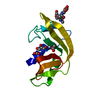
[English] 日本語
Yorodumi
- PDB-3vtt: High Resolution crystal structure of Dengue 3 Envelope protein do... -

+
Open data


ID or keywords:

Loading...

-
Basic information

Entry
Database: PDB / ID: 3vtt
TitleHigh Resolution crystal structure of Dengue 3 Envelope protein domain III (ED3)
ComponentsEnvelope protein E
KeywordsVIRAL PROTEIN / STRUCTURAL PROTEIN / immunoglobin like domain / Epitope presentation / cellular attachment
Function / homology
Function and homology information


symbiont-mediated suppression of host JAK-STAT cascade via inhibition of host TYK2 activity / flavivirin / host cell mitochondrion / symbiont-mediated suppression of host JAK-STAT cascade via inhibition of STAT2 activity / symbiont-mediated suppression of host cytoplasmic pattern recognition receptor signaling pathway via inhibition of MAVS activity / : / viral capsid / nucleoside-triphosphate phosphatase / double-stranded RNA binding / protein complex oligomerization ...symbiont-mediated suppression of host JAK-STAT cascade via inhibition of host TYK2 activity / flavivirin / host cell mitochondrion / symbiont-mediated suppression of host JAK-STAT cascade via inhibition of STAT2 activity / symbiont-mediated suppression of host cytoplasmic pattern recognition receptor signaling pathway via inhibition of MAVS activity / : / viral capsid / nucleoside-triphosphate phosphatase / double-stranded RNA binding / protein complex oligomerization / monoatomic ion channel activity / mRNA (guanine-N7)-methyltransferase / methyltransferase cap1 / clathrin-dependent endocytosis of virus by host cell / mRNA (nucleoside-2'-O-)-methyltransferase activity / mRNA 5'-cap (guanine-N7-)-methyltransferase activity / RNA helicase activity / host cell endoplasmic reticulum membrane / host cell perinuclear region of cytoplasm / protein dimerization activity / RNA helicase / induction by virus of host autophagy / RNA-directed RNA polymerase / viral RNA genome replication / RNA-dependent RNA polymerase activity / serine-type endopeptidase activity / fusion of virus membrane with host endosome membrane / viral envelope / symbiont-mediated suppression of host type I interferon-mediated signaling pathway / host cell nucleus / virion attachment to host cell / virion membrane / structural molecule activity / ATP hydrolysis activity / proteolysis / extracellular region / ATP binding / membrane / metal ion binding
Similarity search - Function
Immunoglobulin-like - #350 / : / Flavivirus envelope glycoprotein E, stem/anchor domain / RNA-directed RNA polymerase, thumb domain, Flavivirus / Flavivirus RNA-directed RNA polymerase, thumb domain / Flavivirus capsid protein C superfamily / : / Flavivirus non-structural protein NS2B / Flavivirus NS3 helicase, C-terminal helical domain / Genome polyprotein, Flavivirus ...Immunoglobulin-like - #350 / : / Flavivirus envelope glycoprotein E, stem/anchor domain / RNA-directed RNA polymerase, thumb domain, Flavivirus / Flavivirus RNA-directed RNA polymerase, thumb domain / Flavivirus capsid protein C superfamily / : / Flavivirus non-structural protein NS2B / Flavivirus NS3 helicase, C-terminal helical domain / Genome polyprotein, Flavivirus / Flavivirus non-structural protein NS4A / Flavivirus non-structural protein NS2B / Flavivirus capsid protein C / Flavivirus non-structural protein NS4B / mRNA cap 0/1 methyltransferase / Flavivirus capsid protein C / Flavivirus non-structural protein NS4B / Flavivirus non-structural protein NS4A / Flavivirus NS2B domain profile. / mRNA cap 0 and cap 1 methyltransferase (EC 2.1.1.56 and EC 2.1.1.57) domain profile. / Flavivirus non-structural protein NS2A / Flavivirus non-structural protein NS2A / Flavivirus NS3, petidase S7 / Peptidase S7, Flavivirus NS3 serine protease / Flavivirus NS3 protease (NS3pro) domain profile. / Envelope glycoprotein M, flavivirus / Flavivirus envelope glycoprotein M / RNA-directed RNA polymerase, flavivirus / Flavivirus RNA-directed RNA polymerase, fingers and palm domains / Flavivirus non-structural Protein NS1 / Flavivirus non-structural protein NS1 / Envelope glycoprotein M superfamily, flavivirus / Flavivirus polyprotein propeptide / Flavivirus polyprotein propeptide superfamily / Flavivirus polyprotein propeptide / Flaviviral glycoprotein E, central domain, subdomain 1 / Flaviviral glycoprotein E, central domain, subdomain 2 / Flavivirus envelope glycoprotein E, Stem/Anchor domain / Flavivirus glycoprotein E, immunoglobulin-like domain / Flavivirus envelope glycoprotein E, Stem/Anchor domain superfamily / Flavivirus glycoprotein, immunoglobulin-like domain / Flavivirus glycoprotein central and dimerisation domain / Flavivirus glycoprotein, central and dimerisation domains / Ribosomal RNA methyltransferase, FtsJ domain / FtsJ-like methyltransferase / Flavivirus/Alphavirus glycoprotein, immunoglobulin-like domain superfamily / Flavivirus glycoprotein, central and dimerisation domain superfamily / Flaviviral glycoprotein E, dimerisation domain / DEAD box, Flavivirus / Flavivirus DEAD domain / helicase superfamily c-terminal domain / Immunoglobulin E-set / Superfamilies 1 and 2 helicase C-terminal domain profile. / Superfamilies 1 and 2 helicase ATP-binding type-1 domain profile. / DEAD-like helicases superfamily / Helicase, C-terminal / Helicase superfamily 1/2, ATP-binding domain / RNA-directed RNA polymerase, catalytic domain / RdRp of positive ssRNA viruses catalytic domain profile. / S-adenosyl-L-methionine-dependent methyltransferase superfamily / Peptidase S1, PA clan, chymotrypsin-like fold / Peptidase S1, PA clan / DNA/RNA polymerase superfamily / Immunoglobulin-like / Sandwich / P-loop containing nucleoside triphosphate hydrolase / Mainly Beta
Similarity search - Domain/homology
Biological speciesDengue virus type 3
MethodX-RAY DIFFRACTION / SYNCHROTRON / MOLECULAR REPLACEMENT / Resolution: 1.7 Å
AuthorsElahi, M. / Islam, M.M. / Kuroda, Y.
CitationJournal: Proteins / Year: 2013
Title: High resolution crystal structure of dengue-3 envelope protein domain III suggests possible molecular mechanisms for serospecific antibody recognition
Authors: Elahi, M. / Islam, M.M. / Noguchi, K. / Yohda, M. / Kuroda, Y.
History
DepositionJun 7, 2012Deposition site: PDBJ / Processing site: PDBJ
Revision 1.0Dec 26, 2012Provider: repository / Type: Initial release
Revision 1.1Aug 7, 2013Group: Database references

-
Structure visualization

Structure viewerMolecule:
MolmilJmol/JSmol

Downloads & links

-
Assembly

Deposited unit
A: Envelope protein E
B: Envelope protein E
hetero molecules


Theoretical massNumber of molelcules
Total (without water)23,4394
Polymers23,2472
Non-polymers1922
Water2,630146
1
A: Envelope protein E
hetero molecules


Theoretical massNumber of molelcules
Total (without water)11,7192
Polymers11,6231
Non-polymers961
Water181
TypeNameSymmetry operationNumber
identity operation1_555x,y,z1
MethodPISA
2
B: Envelope protein E
hetero molecules


Theoretical massNumber of molelcules
Total (without water)11,7192
Polymers11,6231
Non-polymers961
Water181
TypeNameSymmetry operationNumber
identity operation1_555x,y,z1
MethodPISA
Unit cell
Length a, b, c (Å)51.03, 37.68, 52.54
Angle α, β, γ (deg.)90.00, 105.03, 90.00
Int Tables number4
Space group name H-MP1211
DetailsTHE BIOLOGICAL ASSEMBLY OF THE DOMAIN IS TO BE INVESTIGATED.

-
Components

#1: Protein Envelope protein E /


Mass: 11623.287 Da / Num. of mol.: 2 / Fragment: UNP residues 574-678
Source method: isolated from a genetically manipulated source
Source: (gene. exp.) Dengue virus type 3 / Strain: Philippines/H87/1956 / Plasmid: pET15b / Production host: Escherichia coli (E. coli) / Strain (production host): JM109 DE3PlysS / References: UniProt: P27915
#2: Chemical ChemComp-SO4 / SULFATE ION / Sulfate


Mass: 96.063 Da / Num. of mol.: 2 / Source method: obtained synthetically / Formula: SO4
#3: Water ChemComp-HOH / water / Water


Mass: 18.015 Da / Num. of mol.: 146 / Source method: isolated from a natural source / Formula: H2O

-
Experimental details

-
Experiment

ExperimentMethod: X-RAY DIFFRACTION / Number of used crystals: 1

-
Sample preparation

CrystalDensity Matthews: 2.1 Å3/Da / Density % sol: 41.39 %
Crystal growTemperature: 293 K / Method: vapor diffusion, hanging drop / pH: 8.5
Details: 30% PEG 4000, 0.2M Lithium sulfate, 0.1M Tris-HCl, pH 8.5, VAPOR DIFFUSION, HANGING DROP, temperature 293K

-
Data collection

DiffractionMean temperature: 95 K
Diffraction sourceSource: SYNCHROTRON / Site: Photon Factory / Beamline: BL-1A / Wavelength: 1.1 Å
DetectorType: DECTRIS PILATUS 2M / Detector: PIXEL / Date: May 16, 2012
RadiationMonochromator: Si 111 CHANNEL / Protocol: SINGLE WAVELENGTH / Monochromatic (M) / Laue (L): M / Scattering type: x-ray
Radiation wavelengthWavelength: 1.1 Å / Relative weight: 1
ReflectionResolution: 1.7→30.255 Å / Num. all: 21277 / Num. obs: 21522 / % possible obs: 99 % / Observed criterion σ(F): 1.37 / Observed criterion σ(I): 1.37 / Redundancy: 2.8 % / Biso Wilson estimate: 23.64 Å2 / Rmerge(I) obs: 0.038 / Rsym value: 0.034 / Net I/σ(I): 2.31

-
Processing

Software
NameVersionClassification
HKL-2000data collection
MOLREPAuto MRphasing
PHENIX(phenix.refine: 1.7.1_743)refinement
DENZOdata reduction
SCALEPACKdata scaling
RefinementMethod to determine structure: MOLECULAR REPLACEMENT / Resolution: 1.7→30.255 Å / Occupancy max: 1 / Occupancy min: 0.45 / FOM work R set: 0.8328 / SU ML: 0.39 / σ(F): 1.37 / Phase error: 24.14 / Stereochemistry target values: ML
RfactorNum. reflection% reflection
Rfree0.2554 1092 5.14 %
Rwork0.2057 --
obs0.2083 21261 99.01 %
all-21522 -
Solvent computationShrinkage radii: 0.83 Å / VDW probe radii: 1.1 Å / Solvent model: FLAT BULK SOLVENT MODEL / Bsol: 36.257 Å2 / ksol: 0.356 e/Å3
Displacement parametersBiso max: 85.88 Å2 / Biso mean: 33.1108 Å2 / Biso min: 10.53 Å2
Baniso -1Baniso -2Baniso -3
1--0.1565 Å20 Å2-0.1208 Å2
2--0.4109 Å20 Å2
3----0.2544 Å2
Refinement stepCycle: LAST / Resolution: 1.7→30.255 Å
ProteinNucleic acidLigandSolventTotal
Num. atoms1559 0 10 146 1715
Refine LS restraints
Refine-IDTypeDev idealNumber
X-RAY DIFFRACTIONf_bond_d0.0071611
X-RAY DIFFRACTIONf_angle_d1.1452176
X-RAY DIFFRACTIONf_dihedral_angle_d13.998608
X-RAY DIFFRACTIONf_chiral_restr0.077242
X-RAY DIFFRACTIONf_plane_restr0.004279
LS refinement shell

Refine-ID: X-RAY DIFFRACTION / Total num. of bins used: 8

Resolution (Å)Rfactor RfreeNum. reflection RfreeRfactor RworkNum. reflection RworkNum. reflection all% reflection obs (%)
1.7002-1.77760.35911400.28452438257898
1.7776-1.87130.28641340.239325342668100
1.8713-1.98860.28251270.2192512263999
1.9886-2.14210.24761370.214825182655100
2.1421-2.35750.2711410.21332535267699
2.3575-2.69850.27181280.21472529265799
2.6985-3.39910.23281460.20472535268199
3.3991-30.25970.24141390.18442568270798
Refinement TLS params.Method: refined / Origin x: -18.3256 Å / Origin y: 0.0971 Å / Origin z: 14.4097 Å
111213212223313233
T0.1364 Å20.056 Å20.0212 Å2-0.1621 Å20.0477 Å2--0.1541 Å2
L0.1377 °20.0485 °2-0.1146 °2-1.7336 °20.1375 °2--0.2941 °2
S0.0352 Å °0.0201 Å °0.0317 Å °-0.201 Å °-0.0951 Å °-0.291 Å °0.0408 Å °0.0348 Å °0.024 Å °
Refinement TLS groupSelection details: all

+
About Yorodumi

-
News

-
Feb 9, 2022. New format data for meta-information of EMDB entries

New format data for meta-information of EMDB entries

  • Version 3 of the EMDB header file is now the official format.
  • The previous official version 1.9 will be removed from the archive.

Related info.:EMDB header

External links:wwPDB to switch to version 3 of the EMDB data model

-
Aug 12, 2020. Covid-19 info

Covid-19 info

URL: https://pdbj.org/emnavi/covid19.php

New page: Covid-19 featured information page in EM Navigator.

Related info.:Covid-19 info / Mar 5, 2020. Novel coronavirus structure data

+
Mar 5, 2020. Novel coronavirus structure data

Novel coronavirus structure data

Related info.:Yorodumi Speices / Aug 12, 2020. Covid-19 info

External links:COVID-19 featured content - PDBj / Molecule of the Month (242):Coronavirus Proteases

+
Jan 31, 2019. EMDB accession codes are about to change! (news from PDBe EMDB page)

EMDB accession codes are about to change! (news from PDBe EMDB page)

  • The allocation of 4 digits for EMDB accession codes will soon come to an end. Whilst these codes will remain in use, new EMDB accession codes will include an additional digit and will expand incrementally as the available range of codes is exhausted. The current 4-digit format prefixed with “EMD-” (i.e. EMD-XXXX) will advance to a 5-digit format (i.e. EMD-XXXXX), and so on. It is currently estimated that the 4-digit codes will be depleted around Spring 2019, at which point the 5-digit format will come into force.
  • The EM Navigator/Yorodumi systems omit the EMD- prefix.

Related info.:Q: What is EMD? / ID/Accession-code notation in Yorodumi/EM Navigator

External links:EMDB Accession Codes are Changing Soon! / Contact to PDBj

+
Jul 12, 2017. Major update of PDB

Major update of PDB

  • wwPDB released updated PDB data conforming to the new PDBx/mmCIF dictionary.
  • This is a major update changing the version number from 4 to 5, and with Remediation, in which all the entries are updated.
  • In this update, many items about electron microscopy experimental information are reorganized (e.g. em_software).
  • Now, EM Navigator and Yorodumi are based on the updated data.

External links:wwPDB Remediation / Enriched Model Files Conforming to OneDep Data Standards Now Available in the PDB FTP Archive

-
Yorodumi

Thousand views of thousand structures

  • Yorodumi is a browser for structure data from EMDB, PDB, SASBDB, etc.
  • This page is also the successor to EM Navigator detail page, and also detail information page/front-end page for Omokage search.
  • The word "yorodu" (or yorozu) is an old Japanese word meaning "ten thousand". "mi" (miru) is to see.

Related info.:EMDB / PDB / SASBDB / Comparison of 3 databanks / Yorodumi Search / Aug 31, 2016. New EM Navigator & Yorodumi / Yorodumi Papers / Jmol/JSmol / Function and homology information / Changes in new EM Navigator and Yorodumi

Read more