 Movie
Movie Controller


+ Open data

- Basic information
| Entry | Database: PDB / ID: 3up5 | ||||||
|---|---|---|---|---|---|---|---|
| Title | Crystal Structure of OTEMO complex with FAD and NADP (form 4) | ||||||
 Components Components | OTEMO (2,2,3-Trimethyl-5-oxocyclopent-3-enyl)acetyl-CoA 1,5-monooxygenase (2,2,3-Trimethyl-5-oxocyclopent-3-enyl)acetyl-CoA 1,5-monooxygenase | ||||||
 Keywords Keywords | OXIDOREDUCTASE / Baeyer-Villiger monooxygenase | ||||||
| Function / homology |  Function and homology information(2,2,3-trimethyl-5-oxocyclopent-3-enyl)acetyl-CoA 1,5-monooxygenase / (+)-camphor catabolic process / FAD binding / monooxygenase activity / NADP binding / protein homodimerization activity / identical protein binding Function and homology information(2,2,3-trimethyl-5-oxocyclopent-3-enyl)acetyl-CoA 1,5-monooxygenase / (+)-camphor catabolic process / FAD binding / monooxygenase activity / NADP binding / protein homodimerization activity / identical protein bindingSimilarity search - Function | ||||||
| Biological species |  Pseudomonas putida (bacteria) Pseudomonas putida (bacteria) | ||||||
| Method | X-RAY DIFFRACTION / SYNCHROTRON / MOLECULAR REPLACEMENT / Resolution: 2.453 Å | ||||||
 Authors Authors | Shi, R. / Matte, A. / Cygler, M. / Lau, P. | ||||||
 Citation Citation | Journal: Appl.Environ.Microbiol. / Year: 2012 Title: Cloning, Baeyer-Villiger biooxidations, and structures of the camphor pathway 2-oxo-{Delta}(3)-4,5,5-trimethylcyclopentenylacetyl-coenzyme A monooxygenase of Pseudomonas putida ATCC 17453. Authors: Leisch, H. / Shi, R. / Grosse, S. / Morley, K. / Bergeron, H. / Cygler, M. / Iwaki, H. / Hasegawa, Y. / Lau, P.C. | ||||||
| History |
|
- Structure visualization
| Structure viewer | Molecule: MolmilJmol/JSmol |
|---|
- Downloads & links
-Download
| PDBx/mmCIF format | 3up5.cif.gz | 231.6 KB | Display | PDBx/mmCIF format |
|---|---|---|---|---|
| PDB format | pdb3up5.ent.gz | 184.1 KB | Display | PDB format |
| PDBx/mmJSON format | 3up5.json.gz | Tree view | PDBx/mmJSON format | |
| Others |  Other downloads Other downloads |
-Validation report
| Arichive directory | https://data.pdbj.org/pub/pdb/validation_reports/up/3up5ftp://data.pdbj.org/pub/pdb/validation_reports/up/3up5 | HTTPS FTP |
|---|
-Related structure data
| Related structure data |  3uovC  3uoxC  3uoySC  3uozC  3up4C C: citing same article ( S: Starting model for refinement |
|---|---|
| Similar structure data |


-Links
 PDBj
PDBj


- Assembly
| Deposited unit | 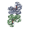
| ||||||||
|---|---|---|---|---|---|---|---|---|---|
| 1 | 
| ||||||||
| 2 | 
| ||||||||
| 3 |
| ||||||||
| Unit cell |
|
-Components
| #1: Protein | (2,2,3-Trimethyl-5-oxocyclopent-3-enyl)acetyl-CoA 1,5-monooxygenase Mass: 61445.949 Da / Num. of mol.: 2 Source method: isolated from a genetically manipulated source Source: (gene. exp.) Pseudomonas putida (bacteria) / Strain: ATCC 17453 / Gene: otemo / Plasmid: pSD80 / Production host: Escherichia coli (E. coli) / Strain (production host): BL21(DE3) / References: UniProt: H3JQW0*PLUS, Oxidoreductases#2: Chemical | Flavin adenine dinucleotide  Mass: 785.550 Da / Num. of mol.: 2 / Source method: obtained synthetically / Formula: C27H33N9O15P2 / Comment: FAD*YM Mass: 785.550 Da / Num. of mol.: 2 / Source method: obtained synthetically / Formula: C27H33N9O15P2 / Comment: FAD*YM#3: Chemical | Nicotinamide adenine dinucleotide phosphate  Mass: 743.405 Da / Num. of mol.: 2 / Source method: obtained synthetically / Formula: C21H28N7O17P3 Mass: 743.405 Da / Num. of mol.: 2 / Source method: obtained synthetically / Formula: C21H28N7O17P3#4: Water | ChemComp-HOH / | Water Mass: 18.015 Da / Num. of mol.: 298 / Source method: isolated from a natural source / Formula: H2O Mass: 18.015 Da / Num. of mol.: 298 / Source method: isolated from a natural source / Formula: H2O |
|---|
-Experimental details
-Experiment
| Experiment | Method: X-RAY DIFFRACTION / Number of used crystals: 1 |
|---|
- Sample preparation
| Crystal | Density Matthews: 2.34 Å3/Da / Density % sol: 47.32 % |
|---|---|
| Crystal grow | Temperature: 295 K / Method: vapor diffusion, sitting drop / pH: 7.5 Details: 22% PEG3350, 0.1 M sodium/potassium phosphate, pH 7.5, VAPOR DIFFUSION, SITTING DROP, temperature 295K |
-Data collection
| Diffraction | Mean temperature: 100 K | |||||||||||||||||||||||||||||||||||||||||||||||||||||||||||||||||||||||||||||
|---|---|---|---|---|---|---|---|---|---|---|---|---|---|---|---|---|---|---|---|---|---|---|---|---|---|---|---|---|---|---|---|---|---|---|---|---|---|---|---|---|---|---|---|---|---|---|---|---|---|---|---|---|---|---|---|---|---|---|---|---|---|---|---|---|---|---|---|---|---|---|---|---|---|---|---|---|---|---|
| Diffraction source | Source: SYNCHROTRON / Site: CLSI  / Beamline: 08ID-1 / Wavelength: 0.9795 Å / Beamline: 08ID-1 / Wavelength: 0.9795 Å | |||||||||||||||||||||||||||||||||||||||||||||||||||||||||||||||||||||||||||||
| Detector | Type: RAYONIX MX-300 / Detector: CCD / Date: Sep 20, 2010 | |||||||||||||||||||||||||||||||||||||||||||||||||||||||||||||||||||||||||||||
| Radiation | Monochromator: double crystal Si(111) / Protocol: SINGLE WAVELENGTH / Monochromatic (M) / Laue (L): M / Scattering type: x-ray | |||||||||||||||||||||||||||||||||||||||||||||||||||||||||||||||||||||||||||||
| Radiation wavelength | Wavelength: 0.9795 Å / Relative weight: 1 | |||||||||||||||||||||||||||||||||||||||||||||||||||||||||||||||||||||||||||||
| Reflection | Resolution: 2.453→90.968 Å / Num. obs: 39964 / % possible obs: 96.6 % / Redundancy: 2.7 % / Rmerge(I) obs: 0.097 / Χ2: 1.49 / Net I/σ(I): 9.2 | |||||||||||||||||||||||||||||||||||||||||||||||||||||||||||||||||||||||||||||
| Reflection shell |
|
- Processing
| Software |
| |||||||||||||||||||||||||||||||||||||||||||||||||||||||||||||||||
|---|---|---|---|---|---|---|---|---|---|---|---|---|---|---|---|---|---|---|---|---|---|---|---|---|---|---|---|---|---|---|---|---|---|---|---|---|---|---|---|---|---|---|---|---|---|---|---|---|---|---|---|---|---|---|---|---|---|---|---|---|---|---|---|---|---|---|
| Refinement | Method to determine structure: MOLECULAR REPLACEMENT Starting model: PDB ENTRY 3UOY Resolution: 2.453→48.353 Å / Cor.coef. Fo:Fc: 0.943 / Cor.coef. Fo:Fc free: 0.899 / WRfactor Rfree: 0.2164 / WRfactor Rwork: 0.1692 / Occupancy max: 1 / Occupancy min: 0.5 / FOM work R set: 0.8431 / SU B: 9.014 / SU ML: 0.203 / SU R Cruickshank DPI: 1.017 / SU Rfree: 0.2909 / Cross valid method: THROUGHOUT / σ(F): 0 / ESU R: 1.017 / ESU R Free: 0.291 / Stereochemistry target values: MAXIMUM LIKELIHOOD Details: HYDROGENS HAVE BEEN ADDED IN THE RIDING POSITIONS U VALUES : REFINED INDIVIDUALLY
| |||||||||||||||||||||||||||||||||||||||||||||||||||||||||||||||||
| Solvent computation | Ion probe radii: 0.8 Å / Shrinkage radii: 0.8 Å / VDW probe radii: 1.4 Å / Solvent model: MASK | |||||||||||||||||||||||||||||||||||||||||||||||||||||||||||||||||
| Displacement parameters | Biso max: 58.26 Å2 / Biso mean: 25.9167 Å2 / Biso min: 8.61 Å2
| |||||||||||||||||||||||||||||||||||||||||||||||||||||||||||||||||
| Refinement step | Cycle: LAST / Resolution: 2.453→48.353 Å
| |||||||||||||||||||||||||||||||||||||||||||||||||||||||||||||||||
| Refine LS restraints |
| |||||||||||||||||||||||||||||||||||||||||||||||||||||||||||||||||
| LS refinement shell | Resolution: 2.453→2.517 Å / Total num. of bins used: 20
|
