 Movie
Movie Controller

[English] 日本語
 Yorodumi
- PDB-3ucg: Crystal structure of a RNA binding domain of polyadenylate-bindin... -
+ Open data

- Basic information
| Entry | Database: PDB / ID: 3ucg | ||||||
|---|---|---|---|---|---|---|---|
| Title | Crystal structure of a RNA binding domain of polyadenylate-binding protein (PABPN1) from Homo sapiens at 1.95 A resolution | ||||||
 Components Components | Polyadenylate-binding protein 2 | ||||||
 Keywords Keywords |  RNA BINDING PROTEIN / Ferredoxin-like / Structural Genomics / Joint Center for Structural Genomics / JCSG / Protein Structure Initiative / PSI-BIOLOGY / Partnership for T-Cell Biology / TCELL RNA BINDING PROTEIN / Ferredoxin-like / Structural Genomics / Joint Center for Structural Genomics / JCSG / Protein Structure Initiative / PSI-BIOLOGY / Partnership for T-Cell Biology / TCELL | ||||||
| Function / homology |  Function and homology information Function and homology informationpositive regulation of polynucleotide adenylyltransferase activity / Inhibition of Host mRNA Processing and RNA Silencing / Processing of Intronless Pre-mRNAs / Z-decay: degradation of maternal mRNAs by zygotically expressed factors / nuclear inclusion body / poly(A) binding / mRNA 3'-end processing / RNA polymerase binding / RNA Polymerase II Transcription Termination / poly(A)+ mRNA export from nucleus ...positive regulation of polynucleotide adenylyltransferase activity / Inhibition of Host mRNA Processing and RNA Silencing / Processing of Intronless Pre-mRNAs / Z-decay: degradation of maternal mRNAs by zygotically expressed factors / nuclear inclusion body / poly(A) binding / mRNA 3'-end processing / RNA polymerase binding / RNA Polymerase II Transcription Termination / poly(A)+ mRNA export from nucleus / Processing of Capped Intron-Containing Pre-mRNA / RNA processing / muscle contraction / mRNA processing / MAPK cascade / cellular response to lipopolysaccharide / nuclear speck / ribonucleoprotein complex / RNA binding / nucleoplasm / nucleus / cytoplasmSimilarity search - Function | ||||||
| Biological species |  Homo sapiens (human) Homo sapiens (human) | ||||||
| Method | X-RAY DIFFRACTION / SYNCHROTRON / MAD / Resolution: 1.95 Å | ||||||
 Authors Authors | Joint Center for Structural Genomics (JCSG) / Partnership for T-Cell Biology (TCELL) | ||||||
 Citation Citation | Journal: To be published Title: Crystal structure of a RNA binding domain of Hypothetical POLYADENYLATE-BINDING PROTEIN (PABPN1) from Homo sapiens at 1.95 A resolution Authors: Joint Center for Structural Genomics (JCSG) / Partnership for T-Cell Biology | ||||||
| History |
|
- Structure visualization
| Structure viewer | Molecule: MolmilJmol/JSmol |
|---|
- Downloads & links
-Download
| PDBx/mmCIF format | 3ucg.cif.gz | 50 KB | Display | PDBx/mmCIF format |
|---|---|---|---|---|
| PDB format | pdb3ucg.ent.gz | 38.8 KB | Display | PDB format |
| PDBx/mmJSON format | 3ucg.json.gz | Tree view | PDBx/mmJSON format | |
| Others |  Other downloads Other downloads |
-Validation report
| Arichive directory | https://data.pdbj.org/pub/pdb/validation_reports/uc/3ucgftp://data.pdbj.org/pub/pdb/validation_reports/uc/3ucg | HTTPS FTP |
|---|
-Related structure data
| Similar structure data | |
|---|---|
| Other databases |


-Links
 PDBj
PDBj- Assembly
| Deposited unit | 
| ||||||||
|---|---|---|---|---|---|---|---|---|---|
| 1 | 
| ||||||||
| Unit cell |
| ||||||||
| Details | PACKING ANALYSIS SUGGEST THE ASSIGNMENT OF A DIMER AS THE SIGNIFICANT OLIGOMERIZATION STATE IN SOLUTION. |
-Components
-Protein , 1 types, 1 molecules A
| #1: Protein | Mass: 9893.984 Da / Num. of mol.: 1 / Fragment: RNA binding domain Source method: isolated from a genetically manipulated source Source: (gene. exp.) Homo sapiens (human) / Gene: BC010939, PAB2, PABP2, PABPN1 / Plasmid: SpeedET / Production host:  Escherichia Coli (E. coli) / Strain (production host): HK100 / References: UniProt: Q86U42 Escherichia Coli (E. coli) / Strain (production host): HK100 / References: UniProt: Q86U42 |
|---|
-Non-polymers , 5 types, 62 molecules 


| #2: Chemical | ChemComp-SO4 / Sulfate Mass: 96.063 Da / Num. of mol.: 1 / Source method: obtained synthetically / Formula: SO4 Mass: 96.063 Da / Num. of mol.: 1 / Source method: obtained synthetically / Formula: SO4 | ||||||
|---|---|---|---|---|---|---|---|
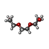
| #3: Chemical | Ethylene glycol Mass: 62.068 Da / Num. of mol.: 2 / Source method: obtained synthetically / Formula: C2H6O2 Mass: 62.068 Da / Num. of mol.: 2 / Source method: obtained synthetically / Formula: C2H6O2#4: Chemical | ChemComp-PEG / | Diethylene glycol Mass: 106.120 Da / Num. of mol.: 1 / Source method: obtained synthetically / Formula: C4H10O3 Mass: 106.120 Da / Num. of mol.: 1 / Source method: obtained synthetically / Formula: C4H10O3#5: Chemical | ChemComp-PGE / | Polyethylene glycol Mass: 150.173 Da / Num. of mol.: 1 / Source method: obtained synthetically / Formula: C6H14O4 Mass: 150.173 Da / Num. of mol.: 1 / Source method: obtained synthetically / Formula: C6H14O4#6: Water | ChemComp-HOH / | WaterMass: 18.015 Da / Num. of mol.: 57 / Source method: isolated from a natural source / Formula: H2O |
-Details
| Sequence details | THIS CONSTRUCT (RESIDUES 167-254) WAS EXPRESSED WITH A PURIFICATION TAG MGSDKIHHHHHHENLYFQG. THE ...THIS CONSTRUCT (RESIDUES 167-254) WAS EXPRESSED WITH A PURIFICATI |
|---|
-Experimental details
-Experiment
| Experiment | Method: X-RAY DIFFRACTION / Number of used crystals: 1 |
|---|
- Sample preparation
| Crystal | Density Matthews: 3.35 Å3/Da / Density % sol: 63.31 % Description: DATA WERE SCALED USING XSCALE WITH FRIEDEL PAIRS KEPT AS SEPARATE WHEN COMPUTING R-SYM, COMPLETENESS AND Crystal grow | Temperature: 277 K / Method: vapor diffusion, sitting drop / pH: 7.5 | Details: 2.0M (NH4)2SO4, 2.0% PEG-400, 0.1M HEPES pH 7.5, NANODROP, VAPOR DIFFUSION, SITTING DROP, temperature 277K |
|---|
-Data collection
| Diffraction | Mean temperature: 100 K | |||||||||||||||||||||||||||||||||||||||||||||||||||||||||||||||||||||||||||||
|---|---|---|---|---|---|---|---|---|---|---|---|---|---|---|---|---|---|---|---|---|---|---|---|---|---|---|---|---|---|---|---|---|---|---|---|---|---|---|---|---|---|---|---|---|---|---|---|---|---|---|---|---|---|---|---|---|---|---|---|---|---|---|---|---|---|---|---|---|---|---|---|---|---|---|---|---|---|---|
| Diffraction source | Source: SYNCHROTRON / Site: SSRL  / Beamline: BL11-1 / Wavelength: 0.91837,0.97925,0.97894 / Beamline: BL11-1 / Wavelength: 0.91837,0.97925,0.97894 | |||||||||||||||||||||||||||||||||||||||||||||||||||||||||||||||||||||||||||||
| Detector | Type: MARMOSAIC 325 mm CCD / Detector: CCD / Date: Jul 21, 2011 Details: Flat mirror (vertical focusing); single crystal Si(111) bent monochromator (ho rizontal focusing) | |||||||||||||||||||||||||||||||||||||||||||||||||||||||||||||||||||||||||||||
| Radiation | Monochromator: single crystal Si(111) bent / Protocol: MAD / Monochromatic (M) / Laue (L): M / Scattering type: x-ray | |||||||||||||||||||||||||||||||||||||||||||||||||||||||||||||||||||||||||||||
| Radiation wavelength |
| |||||||||||||||||||||||||||||||||||||||||||||||||||||||||||||||||||||||||||||
| Reflection | Resolution: 1.95→29.307 Å / Num. obs: 9771 / % possible obs: 99.2 % / Observed criterion σ(I): -3 / Biso Wilson estimate: 37.166 Å2 / Rmerge(I) obs: 0.045 / Net I/σ(I): 18.08 | |||||||||||||||||||||||||||||||||||||||||||||||||||||||||||||||||||||||||||||
| Reflection shell | Diffraction-ID: 1
|
-Phasing
| Phasing | Method: MAD |
|---|
- Processing
| Software |
| |||||||||||||||||||||||||||||||||||||||||||||||||||||||||||||||||||||||||||||||||||||
|---|---|---|---|---|---|---|---|---|---|---|---|---|---|---|---|---|---|---|---|---|---|---|---|---|---|---|---|---|---|---|---|---|---|---|---|---|---|---|---|---|---|---|---|---|---|---|---|---|---|---|---|---|---|---|---|---|---|---|---|---|---|---|---|---|---|---|---|---|---|---|---|---|---|---|---|---|---|---|---|---|---|---|---|---|---|---|
| Refinement | Method to determine structure: MAD / Resolution: 1.95→29.307 Å / Cor.coef. Fo:Fc: 0.97 / Cor.coef. Fo:Fc free: 0.951 / Occupancy max: 1 / Occupancy min: 0.33 / SU B: 6.485 / SU ML: 0.094 / Cross valid method: THROUGHOUT / σ(F): 0 / ESU R Free: 0.122 Stereochemistry target values: MAXIMUM LIKELIHOOD WITH PHASES Details: 1.HYDROGENS HAVE BEEN ADDED IN THE RIDING POSITIONS. 2.A MET-INHIBITION PROTOCOL WAS USED FOR SELENOMETHIONINE INCORPORATION DURING PROTEIN EXPRESSION. THE OCCUPANCY OF THE SE ATOMS IN THE ...Details: 1.HYDROGENS HAVE BEEN ADDED IN THE RIDING POSITIONS. 2.A MET-INHIBITION PROTOCOL WAS USED FOR SELENOMETHIONINE INCORPORATION DURING PROTEIN EXPRESSION. THE OCCUPANCY OF THE SE ATOMS IN THE MSE RESIDUES WAS REDUCED TO 0.75 FOR THE REDUCED SCATTERING POWER DUE TO PARTIAL S-MET INCORPORATION. 3.ATOM RECORDS CONTAIN SUM OF TLS AND RESIDUAL B FACTORS. ANISOU RECORD CONTAINS SUM OF TLS AND RESIDUAL U FACTORS. 4.WATERS WERE EXCLUDED FROM AUTOMATIC TLS ASSIGNMENT. 5.PEG-400 FRAGMENTS (PEG AND PGE) AND SULFATE (SO4) FROM THE CRYSTALLIZATION SOLUTION AND 1,2-ETHANEDIOL (EDO) FROM THE CRYOPROTECTANT HAVE BEEN MODELED IN THE SOLVENT STRUCTURE.
| |||||||||||||||||||||||||||||||||||||||||||||||||||||||||||||||||||||||||||||||||||||
| Solvent computation | Ion probe radii: 0.8 Å / Shrinkage radii: 0.8 Å / VDW probe radii: 1.2 Å / Solvent model: BABINET MODEL WITH MASK | |||||||||||||||||||||||||||||||||||||||||||||||||||||||||||||||||||||||||||||||||||||
| Displacement parameters | Biso max: 183.12 Å2 / Biso mean: 53.3522 Å2 / Biso min: 29.78 Å2 | |||||||||||||||||||||||||||||||||||||||||||||||||||||||||||||||||||||||||||||||||||||
| Refinement step | Cycle: LAST / Resolution: 1.95→29.307 Å
| |||||||||||||||||||||||||||||||||||||||||||||||||||||||||||||||||||||||||||||||||||||
| Refine LS restraints |
| |||||||||||||||||||||||||||||||||||||||||||||||||||||||||||||||||||||||||||||||||||||
| LS refinement shell | Resolution: 1.951→2.002 Å / Total num. of bins used: 20
| |||||||||||||||||||||||||||||||||||||||||||||||||||||||||||||||||||||||||||||||||||||
| Refinement TLS params. | Method: refined / Origin x: 101.6271 Å / Origin y: 76.7715 Å / Origin z: 51.2785 Å
|
