 Movie
Movie Controller

[English] 日本語
 Yorodumi
- PDB-3uaj: Crystal structure of the envelope glycoprotein ectodomain from de... -
+ Open data

- Basic information
| Entry | Database: PDB / ID: 3uaj | ||||||
|---|---|---|---|---|---|---|---|
| Title | Crystal structure of the envelope glycoprotein ectodomain from dengue virus serotype 4 in complex with the fab fragment of the chimpanzee monoclonal antibody 5H2 | ||||||
 Components Components |
| ||||||
 Keywords Keywords |  VIRAL PROTEIN/IMMUNE SYSTEM / dengue antibody membrane fusion / VIRAL PROTEIN-IMMUNE SYSTEM complex VIRAL PROTEIN/IMMUNE SYSTEM / dengue antibody membrane fusion / VIRAL PROTEIN-IMMUNE SYSTEM complex | ||||||
| Function / homology |  Function and homology information Function and homology informationsymbiont-mediated suppression of host JAK-STAT cascade via inhibition of host TYK2 activity / flavivirin / host cell mitochondrion / symbiont-mediated suppression of host JAK-STAT cascade via inhibition of STAT2 activity / symbiont-mediated suppression of host cytoplasmic pattern recognition receptor signaling pathway via inhibition of MAVS activity / ribonucleoside triphosphate phosphatase activity / : / double-stranded RNA binding / viral capsid / nucleoside-triphosphate phosphatase ...symbiont-mediated suppression of host JAK-STAT cascade via inhibition of host TYK2 activity / flavivirin / host cell mitochondrion / symbiont-mediated suppression of host JAK-STAT cascade via inhibition of STAT2 activity / symbiont-mediated suppression of host cytoplasmic pattern recognition receptor signaling pathway via inhibition of MAVS activity / ribonucleoside triphosphate phosphatase activity / : / double-stranded RNA binding / viral capsid / nucleoside-triphosphate phosphatase / protein complex oligomerization / monoatomic ion channel activity / mRNA (guanine-N7)-methyltransferase / methyltransferase cap1 / clathrin-dependent endocytosis of virus by host cell / mRNA (nucleoside-2'-O-)-methyltransferase activity / mRNA 5'-cap (guanine-N7-)-methyltransferase activity / RNA helicase activity / host cell perinuclear region of cytoplasm / host cell endoplasmic reticulum membrane / protein dimerization activity / symbiont-mediated suppression of host type I interferon-mediated signaling pathway / RNA helicase / induction by virus of host autophagy / RNA-directed RNA polymerase / viral RNA genome replication / RNA-dependent RNA polymerase activity / serine-type endopeptidase activity / fusion of virus membrane with host endosome membrane / viral envelope / host cell nucleus / virion attachment to host cell / virion membrane / structural molecule activity / ATP hydrolysis activity / proteolysis / extracellular region / ATP binding / membrane / metal ion bindingSimilarity search - Function | ||||||
| Biological species |  Dengue virus 4 Dengue virus 4 Pan troglodytes (chimpanzee) Pan troglodytes (chimpanzee) | ||||||
| Method | X-RAY DIFFRACTION / SYNCHROTRON / MOLECULAR REPLACEMENT / molecular replacement / Resolution: 3.232 Å | ||||||
 Authors Authors | Cockburn, J.J.B. / Stura, E.A. / Navarro-Sanchez, M.E. / Rey, F.A. | ||||||
 Citation Citation | Journal: Embo J. / Year: 2012 Title: Structural insights into the neutralization mechanism of a higher primate antibody against dengue virus. Authors: Cockburn, J.J. / Navarro Sanchez, M.E. / Goncalvez, A.P. / Zaitseva, E. / Stura, E.A. / Kikuti, C.M. / Duquerroy, S. / Dussart, P. / Chernomordik, L.V. / Lai, C.J. / Rey, F.A. | ||||||
| History |
|
- Structure visualization
| Structure viewer | Molecule: MolmilJmol/JSmol |
|---|
- Downloads & links
-Download
| PDBx/mmCIF format | 3uaj.cif.gz | 316.4 KB | Display | PDBx/mmCIF format |
|---|---|---|---|---|
| PDB format | pdb3uaj.ent.gz | 252 KB | Display | PDB format |
| PDBx/mmJSON format | 3uaj.json.gz | Tree view | PDBx/mmJSON format | |
| Others |  Other downloads Other downloads |
-Validation report
| Arichive directory | https://data.pdbj.org/pub/pdb/validation_reports/ua/3uajftp://data.pdbj.org/pub/pdb/validation_reports/ua/3uaj | HTTPS FTP |
|---|
-Related structure data
| Related structure data |  3uc0C 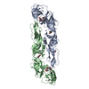 1okeS C: citing same article ( S: Starting model for refinement |
|---|---|
| Similar structure data |


-Links
 PDBj
PDBj


- Assembly
| Deposited unit | 
| ||||||||
|---|---|---|---|---|---|---|---|---|---|
| 1 |
| ||||||||
| Unit cell |
|
-Components
| #1: Protein | Viral envelope Mass: 47285.504 Da / Num. of mol.: 2 / Fragment: UNP residues 280-674 Source method: isolated from a genetically manipulated source Source: (gene. exp.) Dengue virus 4 / Strain: Myanmar 1976 / Gene: envelope / Plasmid: pT351 / Production host:  Drosophila melanogaster (fruit fly) / Strain (production host): Schneider 2 / References: UniProt: Q91AI1, UniProt: P09866*PLUS Drosophila melanogaster (fruit fly) / Strain (production host): Schneider 2 / References: UniProt: Q91AI1, UniProt: P09866*PLUS#2: Antibody | Mass: 25069.979 Da / Num. of mol.: 2 / Fragment: Fab Source method: isolated from a genetically manipulated source Source: (gene. exp.) Pan troglodytes (chimpanzee) / Plasmid: pCOMB3H / Cell line (production host): ovary (CHO) / Production host:  Cricetulus griseus (Chinese hamster) Cricetulus griseus (Chinese hamster)#3: Antibody | Mass: 23493.037 Da / Num. of mol.: 2 / Fragment: Fab Source method: isolated from a genetically manipulated source Source: (gene. exp.) Pan troglodytes (chimpanzee) / Plasmid: pCOMB3H / Cell line (production host): ovary (CHO) / Production host: Cricetulus griseus (Chinese hamster)#4: Sugar | N-Acetylglucosamine  Type: D-saccharide, beta linking / Mass: 221.208 Da / Num. of mol.: 2 Type: D-saccharide, beta linking / Mass: 221.208 Da / Num. of mol.: 2Source method: isolated from a genetically manipulated source Formula: C8H15NO6 |
|---|
-Experimental details
-Experiment
| Experiment | Method: X-RAY DIFFRACTION / Number of used crystals: 1 |
|---|
- Sample preparation
| Crystal | Density Matthews: 3.4 Å3/Da / Density % sol: 63.79 % |
|---|---|
| Crystal grow | Temperature: 292 K / Method: vapor diffusion / pH: 7.5 Details: 8% PEG8000, 8% MPD, 0.1 M HEPES, pH 7.5, VAPOR DIFFUSION, temperature 292K |
-Data collection
| Diffraction | Mean temperature: 100 K | ||||||||||||||||||||||||||||||||||||||||||||||||||||||||||||||||||||||||||||||||||||||||
|---|---|---|---|---|---|---|---|---|---|---|---|---|---|---|---|---|---|---|---|---|---|---|---|---|---|---|---|---|---|---|---|---|---|---|---|---|---|---|---|---|---|---|---|---|---|---|---|---|---|---|---|---|---|---|---|---|---|---|---|---|---|---|---|---|---|---|---|---|---|---|---|---|---|---|---|---|---|---|---|---|---|---|---|---|---|---|---|---|---|
| Diffraction source | Source: SYNCHROTRON / Site: SLS  / Beamline: X06SA / Wavelength: 1 Å / Beamline: X06SA / Wavelength: 1 Å | ||||||||||||||||||||||||||||||||||||||||||||||||||||||||||||||||||||||||||||||||||||||||
| Detector | Type: DECTRIS PILATUS 6M / Detector: PIXEL / Date: May 28, 2008 / Details: Dynamically bendable mirror | ||||||||||||||||||||||||||||||||||||||||||||||||||||||||||||||||||||||||||||||||||||||||
| Radiation | Monochromator: LN2 cooled Fixed-exit Si(111) / Protocol: SINGLE WAVELENGTH / Monochromatic (M) / Laue (L): M / Scattering type: x-ray | ||||||||||||||||||||||||||||||||||||||||||||||||||||||||||||||||||||||||||||||||||||||||
| Radiation wavelength | Wavelength: 1 Å / Relative weight: 1 | ||||||||||||||||||||||||||||||||||||||||||||||||||||||||||||||||||||||||||||||||||||||||
| Reflection | Resolution: 3.232→47.738 Å / Num. all: 41087 / Num. obs: 39891 / % possible obs: 97.3 % / Redundancy: 3.4 % / Biso Wilson estimate: 76.54 Å2 / Rsym value: 0.102 / Net I/σ(I): 10 | ||||||||||||||||||||||||||||||||||||||||||||||||||||||||||||||||||||||||||||||||||||||||
| Reflection shell | Diffraction-ID: 1
|
-Phasing
| Phasing | Method: molecular replacement | |||||||||
|---|---|---|---|---|---|---|---|---|---|---|
| Phasing MR | Rfactor: 54.31 / Model details: Phaser MODE: MR_AUTO
|
- Processing
| Software |
| ||||||||||||||||||||||||||||||||||||||||||||||||||||||||||||||||||||||||||||||||||||||||||||||||||||||||||||
|---|---|---|---|---|---|---|---|---|---|---|---|---|---|---|---|---|---|---|---|---|---|---|---|---|---|---|---|---|---|---|---|---|---|---|---|---|---|---|---|---|---|---|---|---|---|---|---|---|---|---|---|---|---|---|---|---|---|---|---|---|---|---|---|---|---|---|---|---|---|---|---|---|---|---|---|---|---|---|---|---|---|---|---|---|---|---|---|---|---|---|---|---|---|---|---|---|---|---|---|---|---|---|---|---|---|---|---|---|---|
| Refinement | Method to determine structure: MOLECULAR REPLACEMENT Starting model: PDB ENTRY 1OKE Resolution: 3.232→47.738 Å / Cor.coef. Fo:Fc: 0.8535 / Cor.coef. Fo:Fc free: 0.8431 / Occupancy max: 1 / Occupancy min: 1 / Cross valid method: THROUGHOUT / σ(F): 0 / SU Rfree Blow DPI: 0.427 / Stereochemistry target values: Engh & Huber
| ||||||||||||||||||||||||||||||||||||||||||||||||||||||||||||||||||||||||||||||||||||||||||||||||||||||||||||
| Displacement parameters | Biso max: 143.18 Å2 / Biso mean: 69.5232 Å2 / Biso min: 42.04 Å2
| ||||||||||||||||||||||||||||||||||||||||||||||||||||||||||||||||||||||||||||||||||||||||||||||||||||||||||||
| Refine analyze | Luzzati coordinate error obs: 0.77 Å | ||||||||||||||||||||||||||||||||||||||||||||||||||||||||||||||||||||||||||||||||||||||||||||||||||||||||||||
| Refinement step | Cycle: LAST / Resolution: 3.232→47.738 Å
| ||||||||||||||||||||||||||||||||||||||||||||||||||||||||||||||||||||||||||||||||||||||||||||||||||||||||||||
| Refine LS restraints |
| ||||||||||||||||||||||||||||||||||||||||||||||||||||||||||||||||||||||||||||||||||||||||||||||||||||||||||||
| LS refinement shell | Resolution: 3.232→3.31 Å / Total num. of bins used: 20
|
