 Movie
Movie Controller

[English] 日本語
 Yorodumi
- PDB-3tty: Crystal structure of beta-galactosidase from Bacillus circulans s... -
+ Open data

- Basic information
| Entry | Database: PDB / ID: 3tty | ||||||
|---|---|---|---|---|---|---|---|
| Title | Crystal structure of beta-galactosidase from Bacillus circulans sp. alkalophilus in complex with galactose | ||||||
 Components Components | Beta-galactosidase | ||||||
 Keywords Keywords | HYDROLASE / Tim Barrel / Glycoside hydrolase | ||||||
| Function / homology |  Function and homology information Function and homology informationClass I glutamine amidotransferase (GATase) domain / Golgi alpha-mannosidase II / Glycosidases / TIM Barrel / Alpha-Beta Barrel / Immunoglobulin-like / Sandwich / Rossmann fold / 3-Layer(aba) Sandwich / Mainly Beta / Alpha BetaSimilarity search - Domain/homology | ||||||
| Biological species |  Bacillus circulans subsp. alkalophilus (bacteria) Bacillus circulans subsp. alkalophilus (bacteria) | ||||||
| Method | X-RAY DIFFRACTION / SYNCHROTRON / MOLECULAR REPLACEMENT / Resolution: 2.25 Å | ||||||
 Authors Authors | Maksimainen, M. / Hakulinen, N. / Rouvinen, J. | ||||||
 Citation Citation | Journal: Febs J. / Year: 2012 Title: Structural analysis, enzymatic characterization, and catalytic mechanisms of beta-galactosidase from Bacillus circulans sp. alkalophilus. Authors: Maksimainen, M. / Paavilainen, S. / Hakulinen, N. / Rouvinen, J. | ||||||
| History |
|
- Structure visualization
| Structure viewer | Molecule: MolmilJmol/JSmol |
|---|
- Downloads & links
-Download
| PDBx/mmCIF format | 3tty.cif.gz | 839.3 KB | Display | PDBx/mmCIF format |
|---|---|---|---|---|
| PDB format | pdb3tty.ent.gz | 694.6 KB | Display | PDB format |
| PDBx/mmJSON format | 3tty.json.gz | Tree view | PDBx/mmJSON format | |
| Others |  Other downloads Other downloads |
-Validation report
| Arichive directory | https://data.pdbj.org/pub/pdb/validation_reports/tt/3ttyftp://data.pdbj.org/pub/pdb/validation_reports/tt/3tty | HTTPS FTP |
|---|
-Related structure data
| Related structure data |  3ttsSC S: Starting model for refinement C: citing same article ( |
|---|---|
| Similar structure data |


-Links
 PDBj
PDBj- Assembly
| Deposited unit | 
| ||||||||
|---|---|---|---|---|---|---|---|---|---|
| 1 | 
| ||||||||
| 2 | 
| ||||||||
| 3 |
| ||||||||
| Unit cell |
|
-Components
| #1: Protein | / Beta-gal Mass: 77413.359 Da / Num. of mol.: 6 / Source method: isolated from a natural source Source: (natural) Bacillus circulans subsp. alkalophilus (bacteria)References: beta-galactosidase#2: Chemical | ChemComp-ZN /   Mass: 65.409 Da / Num. of mol.: 6 / Source method: obtained synthetically / Formula: Zn Mass: 65.409 Da / Num. of mol.: 6 / Source method: obtained synthetically / Formula: Zn#3: Sugar | ChemComp-GLA / Galactose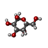  Type: D-saccharide, alpha linking / Mass: 180.156 Da / Num. of mol.: 6 Type: D-saccharide, alpha linking / Mass: 180.156 Da / Num. of mol.: 6Source method: isolated from a genetically manipulated source Formula: C6H12O6 #4: Water | ChemComp-HOH / | Water Mass: 18.015 Da / Num. of mol.: 2516 / Source method: isolated from a natural source / Formula: H2O Mass: 18.015 Da / Num. of mol.: 2516 / Source method: isolated from a natural source / Formula: H2O |
|---|
-Experimental details
-Experiment
| Experiment | Method: X-RAY DIFFRACTION / Number of used crystals: 1 |
|---|
- Sample preparation
| Crystal | Density Matthews: 2.66 Å3/Da / Density % sol: 53.8 % |
|---|---|
| Crystal grow | Temperature: 295 K / Method: vapor diffusion, hanging drop / pH: 8.5 Details: 6-9% PEG 3350, 0.1M magnesium sulfate, 0.1M bicine, pH 8.5, VAPOR DIFFUSION, HANGING DROP, temperature 295K |
-Data collection
| Diffraction | Mean temperature: 100 K |
|---|---|
| Diffraction source | Source: SYNCHROTRON / Site: ESRF  / Beamline: ID14-4 / Wavelength: 0.97625 Å / Beamline: ID14-4 / Wavelength: 0.97625 Å |
| Detector | Type: ADSC QUANTUM 315r / Detector: CCD / Date: Sep 17, 2010 |
| Radiation | Monochromator: Si 111 CHANNEL / Protocol: SINGLE WAVELENGTH / Monochromatic (M) / Laue (L): M / Scattering type: x-ray |
| Radiation wavelength | Wavelength: 0.97625 Å / Relative weight: 1 |
| Reflection | Resolution: 2.25→50 Å / Num. obs: 225561 / % possible obs: 99.2 % / Observed criterion σ(I): 3 / Biso Wilson estimate: 22.18 Å2 |
| Reflection shell | Resolution: 2.25→2.3 Å / % possible all: 97.6 |
- Processing
| Software |
| |||||||||||||||||||||||||||||||||||||||||||||||||||||||||||||||||||||||||||||
|---|---|---|---|---|---|---|---|---|---|---|---|---|---|---|---|---|---|---|---|---|---|---|---|---|---|---|---|---|---|---|---|---|---|---|---|---|---|---|---|---|---|---|---|---|---|---|---|---|---|---|---|---|---|---|---|---|---|---|---|---|---|---|---|---|---|---|---|---|---|---|---|---|---|---|---|---|---|---|
| Refinement | Method to determine structure: MOLECULAR REPLACEMENT Starting model: 3TTS Resolution: 2.25→47.57 Å / SU ML: 0.27 / σ(F): 1.99 / Phase error: 21.49 / Stereochemistry target values: ML
| |||||||||||||||||||||||||||||||||||||||||||||||||||||||||||||||||||||||||||||
| Solvent computation | Shrinkage radii: 0.95 Å / VDW probe radii: 1.2 Å / Solvent model: FLAT BULK SOLVENT MODEL / Bsol: 20.456 Å2 / ksol: 0.319 e/Å3 | |||||||||||||||||||||||||||||||||||||||||||||||||||||||||||||||||||||||||||||
| Displacement parameters |
| |||||||||||||||||||||||||||||||||||||||||||||||||||||||||||||||||||||||||||||
| Refinement step | Cycle: LAST / Resolution: 2.25→47.57 Å
| |||||||||||||||||||||||||||||||||||||||||||||||||||||||||||||||||||||||||||||
| Refine LS restraints |
| |||||||||||||||||||||||||||||||||||||||||||||||||||||||||||||||||||||||||||||
| LS refinement shell |
|
