 Movie
Movie Controller

[English] 日本語
 Yorodumi
- PDB-3tbg: Human cytochrome P450 2D6 with two thioridazines bound in active site -
+ Open data

- Basic information
| Entry | Database: PDB / ID: 3tbg | ||||||
|---|---|---|---|---|---|---|---|
| Title | Human cytochrome P450 2D6 with two thioridazines bound in active site | ||||||
 Components Components | Cytochrome P450 2D6 CYP2D6 CYP2D6 | ||||||
 Keywords Keywords | OXIDOREDUCTASE / Cytochrome P450 / monooxygenase / thioridazine | ||||||
| Function / homology |  Function and homology information Function and homology informationnegative regulation of binding / negative regulation of cellular organofluorine metabolic process / Miscellaneous substrates / isoquinoline alkaloid metabolic process / Fatty acids / coumarin metabolic process / Oxidoreductases; Acting on paired donors, with incorporation or reduction of molecular oxygen; With reduced flavin or flavoprotein as one donor, and incorporation of one atom of oxygen into the other donor / CYP2E1 reactions / arachidonic acid metabolic process / anandamide 8,9 epoxidase activity ...negative regulation of binding / negative regulation of cellular organofluorine metabolic process / Miscellaneous substrates / isoquinoline alkaloid metabolic process / Fatty acids / coumarin metabolic process / Oxidoreductases; Acting on paired donors, with incorporation or reduction of molecular oxygen; With reduced flavin or flavoprotein as one donor, and incorporation of one atom of oxygen into the other donor / CYP2E1 reactions / arachidonic acid metabolic process / anandamide 8,9 epoxidase activity / anandamide 11,12 epoxidase activity / anandamide 14,15 epoxidase activity / alkaloid catabolic process / alkaloid metabolic process / : / Biosynthesis of maresin-like SPMs / monoterpenoid metabolic process / Xenobiotics / oxidative demethylation / oxidoreductase activity, acting on paired donors, with incorporation or reduction of molecular oxygen, reduced flavin or flavoprotein as one donor, and incorporation of one atom of oxygen / long-chain fatty acid biosynthetic process / estrogen metabolic process / retinol metabolic process / Aspirin ADME / steroid metabolic process / xenobiotic catabolic process / xenobiotic metabolic process / cholesterol metabolic process / monooxygenase activity / oxidoreductase activity / iron ion binding / intracellular membrane-bounded organelle / heme binding / endoplasmic reticulum membrane / endoplasmic reticulum / mitochondrion / cytoplasmSimilarity search - Function | ||||||
| Biological species |  Homo sapiens (human) Homo sapiens (human) | ||||||
| Method | X-RAY DIFFRACTION / SYNCHROTRON / MOLECULAR REPLACEMENT / Resolution: 2.1 Å | ||||||
 Authors Authors | Wang, A. / Stout, C.D. / Johnson, E.F. | ||||||
 Citation Citation | Journal: J.Biol.Chem. / Year: 2015 Title: Contributions of Ionic Interactions and Protein Dynamics to Cytochrome P450 2D6 (CYP2D6) Substrate and Inhibitor Binding. Authors: Wang, A. / Stout, C.D. / Zhang, Q. / Johnson, E.F. | ||||||
| History |
|
- Structure visualization
| Structure viewer | Molecule: MolmilJmol/JSmol |
|---|
- Downloads & links
-Download
| PDBx/mmCIF format | 3tbg.cif.gz | 388.1 KB | Display | PDBx/mmCIF format |
|---|---|---|---|---|
| PDB format | pdb3tbg.ent.gz | 316.6 KB | Display | PDB format |
| PDBx/mmJSON format | 3tbg.json.gz | Tree view | PDBx/mmJSON format | |
| Others |  Other downloads Other downloads |
-Validation report
| Arichive directory | https://data.pdbj.org/pub/pdb/validation_reports/tb/3tbgftp://data.pdbj.org/pub/pdb/validation_reports/tb/3tbg | HTTPS FTP |
|---|
-Related structure data
| Related structure data |  3tdaC  4wntC  4wnuC 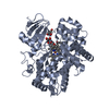 4wnvC 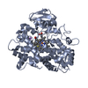 4wnwC  3qm4S S: Starting model for refinement C: citing same article ( |
|---|---|
| Similar structure data |


-Links
 PDBj
PDBj

- Assembly
| Deposited unit | 
| ||||||||
|---|---|---|---|---|---|---|---|---|---|
| 1 | 
| ||||||||
| 2 | 
| ||||||||
| 3 | 
| ||||||||
| 4 | 
| ||||||||
| Unit cell |
|
-Components
-Protein , 1 types, 4 molecules ABCD
| #1: Protein | CYP2D6 / CYPIID6 / Cytochrome P450-DB1 / Debrisoquine 4-hydroxylase Mass: 53730.566 Da / Num. of mol.: 4 / Fragment: UNP residues 34-497 Source method: isolated from a genetically manipulated source Source: (gene. exp.) Homo sapiens (human) / Gene: CYP2D6, CYP2DL1 / Plasmid: pCWORI / Production host:  Escherichia coli (E. coli) / Strain (production host): DH5alpha / References: UniProt: P10635, unspecific monooxygenase Escherichia coli (E. coli) / Strain (production host): DH5alpha / References: UniProt: P10635, unspecific monooxygenase |
|---|
-Non-polymers , 6 types, 531 molecules 


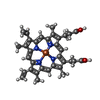

| #2: Chemical | ChemComp-RTZ /  Mass: 370.575 Da / Num. of mol.: 8 / Source method: obtained synthetically / Formula: C21H26N2S2 Mass: 370.575 Da / Num. of mol.: 8 / Source method: obtained synthetically / Formula: C21H26N2S2#3: Chemical | ChemComp-ZN /  Mass: 65.409 Da / Num. of mol.: 7 / Source method: obtained synthetically / Formula: Zn Mass: 65.409 Da / Num. of mol.: 7 / Source method: obtained synthetically / Formula: Zn#4: Chemical | ChemComp-GOL / Glycerol Mass: 92.094 Da / Num. of mol.: 7 / Source method: obtained synthetically / Formula: C3H8O3 Mass: 92.094 Da / Num. of mol.: 7 / Source method: obtained synthetically / Formula: C3H8O3#5: Chemical | ChemComp-PO4 / | Phosphate Mass: 94.971 Da / Num. of mol.: 1 / Source method: obtained synthetically / Formula: PO4 Mass: 94.971 Da / Num. of mol.: 1 / Source method: obtained synthetically / Formula: PO4#6: Chemical | ChemComp-HEM / Heme B Mass: 616.487 Da / Num. of mol.: 4 / Source method: obtained synthetically / Formula: C34H32FeN4O4 Mass: 616.487 Da / Num. of mol.: 4 / Source method: obtained synthetically / Formula: C34H32FeN4O4#7: Water | ChemComp-HOH / | WaterMass: 18.015 Da / Num. of mol.: 504 / Source method: isolated from a natural source / Formula: H2O |
|---|
-Experimental details
-Experiment
| Experiment | Method: X-RAY DIFFRACTION / Number of used crystals: 1 |
|---|
- Sample preparation
| Crystal | Density Matthews: 3.19 Å3/Da / Density % sol: 61.39 % |
|---|---|
| Crystal grow | Temperature: 298 K / Method: vapor diffusion, hanging drop / pH: 7 Details: PEG3350, sodium acetate, sodium cacodylate, potassium phosphate, sodium chloride, zinc chloride, glycerol, beta-mercaptoethanol, thioridazine, HEGA-10, facial amphiphile 231_CHOL, pH 7.0, ...Details: PEG3350, sodium acetate, sodium cacodylate, potassium phosphate, sodium chloride, zinc chloride, glycerol, beta-mercaptoethanol, thioridazine, HEGA-10, facial amphiphile 231_CHOL, pH 7.0, VAPOR DIFFUSION, HANGING DROP, temperature 298K |
-Data collection
| Diffraction | Mean temperature: 100 K |
|---|---|
| Diffraction source | Source: SYNCHROTRON / Site: SSRL  / Beamline: BL7-1 / Wavelength: 0.97945 Å / Beamline: BL7-1 / Wavelength: 0.97945 Å |
| Detector | Type: ADSC QUANTUM 315r / Detector: CCD / Date: Mar 18, 2011 / Details: Rh coated flat mirror |
| Radiation | Monochromator: Side scattering bent cube-root I-beam single crystal Protocol: SINGLE WAVELENGTH / Monochromatic (M) / Laue (L): M / Scattering type: x-ray |
| Radiation wavelength | Wavelength: 0.97945 Å / Relative weight: 1 |
| Reflection | Resolution: 2.04→37.957 Å / Num. all: 174928 / Num. obs: 174399 / % possible obs: 99.8 % / Observed criterion σ(F): 0 / Observed criterion σ(I): 0 / Redundancy: 3.9 % / Biso Wilson estimate: 24.036 Å2 / Rmerge(I) obs: 0.088 / Rsym value: 0.088 / Net I/σ(I): 4.8 |
| Reflection shell | Resolution: 2.04→2.15 Å / Redundancy: 4 % / Rmerge(I) obs: 0.352 / Mean I/σ(I) obs: 1.5 / Num. unique all: 25169 / Rsym value: 0.352 / % possible all: 100 |
- Processing
| Software |
| ||||||||||||||||||||||||||||||||||||||||||||||||||||||||||||||||||||||||||||||||
|---|---|---|---|---|---|---|---|---|---|---|---|---|---|---|---|---|---|---|---|---|---|---|---|---|---|---|---|---|---|---|---|---|---|---|---|---|---|---|---|---|---|---|---|---|---|---|---|---|---|---|---|---|---|---|---|---|---|---|---|---|---|---|---|---|---|---|---|---|---|---|---|---|---|---|---|---|---|---|---|---|---|
| Refinement | Method to determine structure: MOLECULAR REPLACEMENT Starting model: PDB ENTRY 3QM4 Resolution: 2.1→36.9 Å / Rfactor Rfree error: 0.003 / Data cutoff high absF: 4551940.29 / Data cutoff low absF: 0 / Isotropic thermal model: RESTRAINED / Cross valid method: THROUGHOUT / σ(F): 0 / Stereochemistry target values: Engh & Huber / Details: BULK SOLVENT MODEL USED
| ||||||||||||||||||||||||||||||||||||||||||||||||||||||||||||||||||||||||||||||||
| Solvent computation | Solvent model: FLAT MODEL / Bsol: 54.5701 Å2 / ksol: 0.4 e/Å3 | ||||||||||||||||||||||||||||||||||||||||||||||||||||||||||||||||||||||||||||||||
| Displacement parameters | Biso mean: 34.8 Å2
| ||||||||||||||||||||||||||||||||||||||||||||||||||||||||||||||||||||||||||||||||
| Refine analyze |
| ||||||||||||||||||||||||||||||||||||||||||||||||||||||||||||||||||||||||||||||||
| Refinement step | Cycle: LAST / Resolution: 2.1→36.9 Å
| ||||||||||||||||||||||||||||||||||||||||||||||||||||||||||||||||||||||||||||||||
| Refine LS restraints |
| ||||||||||||||||||||||||||||||||||||||||||||||||||||||||||||||||||||||||||||||||
| Refine LS restraints NCS | NCS model details: NONE | ||||||||||||||||||||||||||||||||||||||||||||||||||||||||||||||||||||||||||||||||
| LS refinement shell | Resolution: 2.1→2.23 Å / Rfactor Rfree error: 0.008 / Total num. of bins used: 6
| ||||||||||||||||||||||||||||||||||||||||||||||||||||||||||||||||||||||||||||||||
| Xplor file |
|
