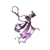
 Movie
Movie Controller

[English] 日本語
 Yorodumi
- PDB-3sgm: Bromoderivative-2 of amyloid-related segment of alphaB-crystallin... -
+ Open data

- Basic information
| Entry | Database: PDB / ID: 3sgm | ||||||
|---|---|---|---|---|---|---|---|
| Title | Bromoderivative-2 of amyloid-related segment of alphaB-crystallin residues 90-100 | ||||||
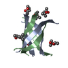
 Components Components | Alpha-crystallin B chain CRYAB CRYAB | ||||||
 Keywords Keywords | PROTEIN FIBRIL / amyloid / amyloid oligomer / beta cylindrin | ||||||
| Function / homology |  Function and homology information Function and homology informationmicrotubule polymerization or depolymerization / negative regulation of intracellular transport / apoptotic process involved in morphogenesis / cardiac myofibril / regulation of programmed cell death / tubulin complex assembly / structural constituent of eye lens / negative regulation of amyloid fibril formation / M band / lens development in camera-type eye ...microtubule polymerization or depolymerization / negative regulation of intracellular transport / apoptotic process involved in morphogenesis / cardiac myofibril / regulation of programmed cell death / tubulin complex assembly / structural constituent of eye lens / negative regulation of amyloid fibril formation / M band / lens development in camera-type eye / muscle organ development / actin filament bundle / HSF1-dependent transactivation / negative regulation of reactive oxygen species metabolic process / negative regulation of protein-containing complex assembly / stress-activated MAPK cascade / muscle contraction / synaptic membrane / response to hydrogen peroxide / cellular response to gamma radiation / negative regulation of cell growth / Z disc / unfolded protein binding / protein folding / response to estradiol / amyloid-beta binding / response to heat / perikaryon / protein refolding / microtubule binding / dendritic spine / lysosome / response to hypoxia / protein stabilization / axon / negative regulation of gene expression / negative regulation of DNA-templated transcription / protein-containing complex binding / negative regulation of apoptotic process / structural molecule activity / cell surface / protein homodimerization activity / protein-containing complex / mitochondrion / extracellular exosome / nucleoplasm / identical protein binding / metal ion binding / nucleus / cytosol / cytoplasmSimilarity search - Function | ||||||
| Biological species |  Homo sapiens (human) Homo sapiens (human) | ||||||
| Method | X-RAY DIFFRACTION / SYNCHROTRON / SAD / MAD / Resolution: 1.7006 Å | ||||||
 Authors Authors | Laganowsky, A. / Sawaya, M.R. / Cascio, D. / Eisenberg, D. | ||||||
 Citation Citation | Journal: Science / Year: 2012 Title: Atomic view of a toxic amyloid small oligomer. Authors: Laganowsky, A. / Liu, C. / Sawaya, M.R. / Whitelegge, J.P. / Park, J. / Zhao, M. / Pensalfini, A. / Soriaga, A.B. / Landau, M. / Teng, P.K. / Cascio, D. / Glabe, C. / Eisenberg, D. | ||||||
| History |
|
- Structure visualization
| Structure viewer | Molecule: MolmilJmol/JSmol |
|---|
- Downloads & links
-Download
| PDBx/mmCIF format | 3sgm.cif.gz | 26.3 KB | Display | PDBx/mmCIF format |
|---|---|---|---|---|
| PDB format | pdb3sgm.ent.gz | 21 KB | Display | PDB format |
| PDBx/mmJSON format | 3sgm.json.gz | Tree view | PDBx/mmJSON format | |
| Others |  Other downloads Other downloads |
-Validation report
| Arichive directory | https://data.pdbj.org/pub/pdb/validation_reports/sg/3sgmftp://data.pdbj.org/pub/pdb/validation_reports/sg/3sgm | HTTPS FTP |
|---|
-Related structure data


-Links
 PDBj
PDBj


- Assembly
| Deposited unit | 
| ||||||||
|---|---|---|---|---|---|---|---|---|---|
| 1 | 
| ||||||||
| 2 | 
| ||||||||
| Unit cell |
|
-Components
| #1: Protein/peptide | CRYAB / Alpha(B)-crystallin / Heat shock protein beta-5 / HspB5 / Renal carcinoma antigen NY-REN-27 / ...Alpha(B)-crystallin / Heat shock protein beta-5 / HspB5 / Renal carcinoma antigen NY-REN-27 / Rosenthal fiber component Mass: 1277.348 Da / Num. of mol.: 4 / Source method: obtained synthetically / Details: synthetic peptide / Source: (synth.) Homo sapiens (human) / References: UniProt: P02511#2: Chemical | ChemComp-MPD / ( | 2-Methyl-2,4-pentanediol  Mass: 118.174 Da / Num. of mol.: 1 / Source method: obtained synthetically / Formula: C6H14O2 / Comment: precipitant*YM Mass: 118.174 Da / Num. of mol.: 1 / Source method: obtained synthetically / Formula: C6H14O2 / Comment: precipitant*YM#3: Water | ChemComp-HOH / | Water Mass: 18.015 Da / Num. of mol.: 16 / Source method: isolated from a natural source / Formula: H2O Mass: 18.015 Da / Num. of mol.: 16 / Source method: isolated from a natural source / Formula: H2O |
|---|
-Experimental details
-Experiment
| Experiment | Method: X-RAY DIFFRACTION / Number of used crystals: 1 |
|---|
- Sample preparation
| Crystal | Density Matthews: 2.34 Å3/Da / Density % sol: 47.52 % |
|---|---|
| Crystal grow | Temperature: 298 K / Method: vapor diffusion, hanging drop / pH: 7 Details: 0.1M TRIS pH 7.0, 35% MPD, 0.2M SODIUM CHLORIDE, vapor diffusion, hanging drop, temperature 298K |
-Data collection
| Diffraction |
| |||||||||||||||||||||||||||||||||||||||||||||||||||||||||||||||||||||||||||||
|---|---|---|---|---|---|---|---|---|---|---|---|---|---|---|---|---|---|---|---|---|---|---|---|---|---|---|---|---|---|---|---|---|---|---|---|---|---|---|---|---|---|---|---|---|---|---|---|---|---|---|---|---|---|---|---|---|---|---|---|---|---|---|---|---|---|---|---|---|---|---|---|---|---|---|---|---|---|---|
| Diffraction source |
| |||||||||||||||||||||||||||||||||||||||||||||||||||||||||||||||||||||||||||||
| Detector |
| |||||||||||||||||||||||||||||||||||||||||||||||||||||||||||||||||||||||||||||
| Radiation |
| |||||||||||||||||||||||||||||||||||||||||||||||||||||||||||||||||||||||||||||
| Radiation wavelength |
| |||||||||||||||||||||||||||||||||||||||||||||||||||||||||||||||||||||||||||||
| Reflection | Resolution: 1.62→50 Å / Num. obs: 6043 / % possible obs: 97.2 % / Redundancy: 3.6 % / Rmerge(I) obs: 0.033 / Χ2: 1.004 / Net I/σ(I): 28.6 | |||||||||||||||||||||||||||||||||||||||||||||||||||||||||||||||||||||||||||||
| Reflection shell |
|

-Phasing
| Phasing | Method: MAD |
|---|
- Processing
| Software |
| ||||||||||||||||||||||||||||||||
|---|---|---|---|---|---|---|---|---|---|---|---|---|---|---|---|---|---|---|---|---|---|---|---|---|---|---|---|---|---|---|---|---|---|
| Refinement | Method to determine structure: SAD / Resolution: 1.7006→32.998 Å / Occupancy max: 1 / Occupancy min: 0.35 / FOM work R set: 0.8159 / SU ML: 0.32 / σ(F): 1.41 / Phase error: 24.29 / Stereochemistry target values: ML
| ||||||||||||||||||||||||||||||||
| Solvent computation | Shrinkage radii: 0.83 Å / VDW probe radii: 1.1 Å / Solvent model: FLAT BULK SOLVENT MODEL / Bsol: 60.966 Å2 / ksol: 0.43 e/Å3 | ||||||||||||||||||||||||||||||||
| Displacement parameters | Biso max: 43.96 Å2 / Biso mean: 21.8548 Å2 / Biso min: 10.47 Å2
| ||||||||||||||||||||||||||||||||
| Refinement step | Cycle: LAST / Resolution: 1.7006→32.998 Å
| ||||||||||||||||||||||||||||||||
| Refine LS restraints |
| ||||||||||||||||||||||||||||||||
| LS refinement shell | Refine-ID: X-RAY DIFFRACTION / Total num. of bins used: 2 / % reflection obs: 100 %
|
