 Movie
Movie Controller

[English] 日本語
 Yorodumi
- PDB-3q4t: Crystal structure of Activin receptor type-IIA (ACVR2A) kinase do... -
+ Open data

- Basic information
| Entry | Database: PDB / ID: 3q4t | ||||||
|---|---|---|---|---|---|---|---|
| Title | Crystal structure of Activin receptor type-IIA (ACVR2A) kinase domain in complex with dorsomorphin | ||||||
 Components Components | Activin receptor type-2A | ||||||
 Keywords Keywords |  TRANSFERASE / Structural Genomics Consortium / SGC / Protein kinase TRANSFERASE / Structural Genomics Consortium / SGC / Protein kinase | ||||||
| Function / homology |  Function and homology information Function and homology informationRegulation of signaling by NODAL / inhibin-betaglycan-ActRII complex / inhibin binding / penile erection / positive regulation of activin receptor signaling pathway / activin receptor activity / Sertoli cell proliferation / sperm ejaculation / BMP receptor activity / embryonic skeletal system development ...Regulation of signaling by NODAL / inhibin-betaglycan-ActRII complex / inhibin binding / penile erection / positive regulation of activin receptor signaling pathway / activin receptor activity / Sertoli cell proliferation / sperm ejaculation / BMP receptor activity / embryonic skeletal system development / activin receptor complex / receptor protein serine/threonine kinase / transmembrane receptor protein serine/threonine kinase activity / Signaling by BMP / activin binding / cellular response to BMP stimulus / activin receptor signaling pathway / Signaling by Activin / Signaling by NODAL / gastrulation with mouth forming second / regulation of nitric oxide biosynthetic process / determination of left/right symmetry / anterior/posterior pattern specification / cell surface receptor protein serine/threonine kinase signaling pathway / odontogenesis of dentin-containing tooth / growth factor binding / mesoderm development / positive regulation of SMAD protein signal transduction / BMP signaling pathway / positive regulation of bone mineralization / positive regulation of osteoblast differentiation / coreceptor activity / positive regulation of erythrocyte differentiation / PDZ domain binding / cellular response to growth factor stimulus / : / spermatogenesis / receptor complex / positive regulation of protein phosphorylation / phosphorylation / protein serine/threonine kinase activity / cell surface / positive regulation of transcription by RNA polymerase II / ATP binding / metal ion binding / plasma membrane / cytoplasmSimilarity search - Function | ||||||
| Biological species |  Homo sapiens (human) Homo sapiens (human) | ||||||
| Method | X-RAY DIFFRACTION / SYNCHROTRON / MOLECULAR REPLACEMENT / Resolution: 1.96 Å | ||||||
 Authors Authors | Chaikuad, A. / Alfano, I. / Mahajan, P. / Cooper, C.D.O. / Sanvitale, C. / Vollmar, M. / Krojer, T. / Muniz, J.R.C. / Raynor, J. / von Delft, F. ...Chaikuad, A. / Alfano, I. / Mahajan, P. / Cooper, C.D.O. / Sanvitale, C. / Vollmar, M. / Krojer, T. / Muniz, J.R.C. / Raynor, J. / von Delft, F. / Weigelt, J. / Arrowsmith, C.H. / Edwards, A.M. / Bountra, C. / Bullock, A. / Structural Genomics Consortium (SGC) | ||||||
 Citation Citation | Journal: J.Biol.Chem. / Year: 2015 Title: Small Molecules Dorsomorphin and LDN-193189 Inhibit Myostatin/GDF8 Signaling and Promote Functional Myoblast Differentiation. Authors: Horbelt, D. / Boergermann, J.H. / Chaikuad, A. / Alfano, I. / Williams, E. / Lukonin, I. / Timmel, T. / Bullock, A.N. / Knaus, P. | ||||||
| History |
|
- Structure visualization
| Structure viewer | Molecule: MolmilJmol/JSmol |
|---|
- Downloads & links
-Download
| PDBx/mmCIF format | 3q4t.cif.gz | 270.7 KB | Display | PDBx/mmCIF format |
|---|---|---|---|---|
| PDB format | pdb3q4t.ent.gz | 218.8 KB | Display | PDB format |
| PDBx/mmJSON format | 3q4t.json.gz | Tree view | PDBx/mmJSON format | |
| Others |  Other downloads Other downloads |
-Validation report
| Arichive directory | https://data.pdbj.org/pub/pdb/validation_reports/q4/3q4tftp://data.pdbj.org/pub/pdb/validation_reports/q4/3q4t | HTTPS FTP |
|---|
-Related structure data
| Related structure data |  2qluS S: Starting model for refinement |
|---|---|
| Similar structure data |


-Links
 PDBj
PDBj

- Assembly
| Deposited unit | 
| ||||||||
|---|---|---|---|---|---|---|---|---|---|
| 1 | 
| ||||||||
| 2 | 
| ||||||||
| Unit cell |
|
-Components
-Protein , 1 types, 2 molecules AB
| #1: Protein | Mass: 36548.766 Da / Num. of mol.: 2 / Fragment: kinase domain (UNP residues 191-488) Source method: isolated from a genetically manipulated source Source: (gene. exp.) Homo sapiens (human) / Gene: ACVR2, ACVR2A / Plasmid: pFB-LIC-Bse / Cell line (production host): SF9 / Production host:   Spodoptera frugiperda (fall armyworm) Spodoptera frugiperda (fall armyworm)References: UniProt: P27037, receptor protein serine/threonine kinase |
|---|
-Non-polymers , 5 types, 524 molecules 


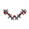

| #2: Chemical |  Mass: 399.488 Da / Num. of mol.: 2 / Source method: obtained synthetically / Formula: C24H25N5O Mass: 399.488 Da / Num. of mol.: 2 / Source method: obtained synthetically / Formula: C24H25N5O#3: Chemical | ChemComp-SO4 / Sulfate Mass: 96.063 Da / Num. of mol.: 4 / Source method: obtained synthetically / Formula: SO4 Mass: 96.063 Da / Num. of mol.: 4 / Source method: obtained synthetically / Formula: SO4#4: Chemical | ChemComp-EDO / Ethylene glycol Mass: 62.068 Da / Num. of mol.: 21 / Source method: obtained synthetically / Formula: C2H6O2 Mass: 62.068 Da / Num. of mol.: 21 / Source method: obtained synthetically / Formula: C2H6O2#5: Chemical | ChemComp-PG4 / | Polyethylene glycol Mass: 194.226 Da / Num. of mol.: 1 / Source method: obtained synthetically / Formula: C8H18O5 / Comment: precipitant*YM Mass: 194.226 Da / Num. of mol.: 1 / Source method: obtained synthetically / Formula: C8H18O5 / Comment: precipitant*YM#6: Water | ChemComp-HOH / | WaterMass: 18.015 Da / Num. of mol.: 496 / Source method: isolated from a natural source / Formula: H2O |
|---|
-Experimental details
-Experiment
| Experiment | Method: X-RAY DIFFRACTION / Number of used crystals: 1 |
|---|
- Sample preparation
| Crystal | Density Matthews: 2.48 Å3/Da / Density % sol: 50.33 % |
|---|---|
| Crystal grow | Temperature: 293.15 K / Method: vapor diffusion, sitting drop / pH: 8.8 Details: 28% PEG 3350, 0.2M LiSO4, 0.1M Tris, pH 8.8, 10% Ethylene glycol, VAPOR DIFFUSION, SITTING DROP, temperature 293.15K |
-Data collection
| Diffraction | Mean temperature: 100 K |
|---|---|
| Diffraction source | Source: SYNCHROTRON / Site: Diamond  / Beamline: I03 / Wavelength: 0.9763 Å / Beamline: I03 / Wavelength: 0.9763 Å |
| Detector | Type: ADSC Q315 3x3 CCD / Detector: CCD / Date: Apr 21, 2010 / Details: Kirkpatrick Baez bimorph mirror pair |
| Radiation | Monochromator: Si (111) double crystal monochromator / Protocol: SINGLE WAVELENGTH / Monochromatic (M) / Laue (L): M / Scattering type: x-ray |
| Radiation wavelength | Wavelength: 0.9763 Å / Relative weight: 1 |
| Reflection | Resolution: 1.96→48.6 Å / Num. all: 53883 / Num. obs: 53760 / % possible obs: 99.9 % / Observed criterion σ(F): 0 / Observed criterion σ(I): 0 / Redundancy: 7 % / Biso Wilson estimate: 26.6 Å2 / Rmerge(I) obs: 0.112 / Net I/σ(I): 11.2 |
| Reflection shell | Resolution: 1.96→2.07 Å / Redundancy: 7.2 % / Rmerge(I) obs: 0.783 / Mean I/σ(I) obs: 2.3 / Num. unique all: 7727 / % possible all: 100 |
- Processing
| Software |
| |||||||||||||||||||||||||||||||||||||||||||||||||||||||||||||||||||||||||||||||||||||||||||||||||||||||||||||||||||||||||||||||||||||||||||||||||||||||||||||||||||||||||||||||||||||||||||||||||||||||||||||||||||||||||||||||||
|---|---|---|---|---|---|---|---|---|---|---|---|---|---|---|---|---|---|---|---|---|---|---|---|---|---|---|---|---|---|---|---|---|---|---|---|---|---|---|---|---|---|---|---|---|---|---|---|---|---|---|---|---|---|---|---|---|---|---|---|---|---|---|---|---|---|---|---|---|---|---|---|---|---|---|---|---|---|---|---|---|---|---|---|---|---|---|---|---|---|---|---|---|---|---|---|---|---|---|---|---|---|---|---|---|---|---|---|---|---|---|---|---|---|---|---|---|---|---|---|---|---|---|---|---|---|---|---|---|---|---|---|---|---|---|---|---|---|---|---|---|---|---|---|---|---|---|---|---|---|---|---|---|---|---|---|---|---|---|---|---|---|---|---|---|---|---|---|---|---|---|---|---|---|---|---|---|---|---|---|---|---|---|---|---|---|---|---|---|---|---|---|---|---|---|---|---|---|---|---|---|---|---|---|---|---|---|---|---|---|---|---|---|---|---|---|---|---|---|---|---|---|---|---|---|---|---|
| Refinement | Method to determine structure: MOLECULAR REPLACEMENT Starting model: pdb id 2qlu Resolution: 1.96→45.47 Å / Cor.coef. Fo:Fc: 0.963 / Cor.coef. Fo:Fc free: 0.939 / SU B: 6.75 / SU ML: 0.1 / Cross valid method: THROUGHOUT / σ(F): 0 / σ(I): 2 / ESU R Free: 0.147 / Stereochemistry target values: MAXIMUM LIKELIHOOD Details: HYDROGENS HAVE BEEN USED IN REFINEMENT BUT NOT OUTPUT TO PDB
| |||||||||||||||||||||||||||||||||||||||||||||||||||||||||||||||||||||||||||||||||||||||||||||||||||||||||||||||||||||||||||||||||||||||||||||||||||||||||||||||||||||||||||||||||||||||||||||||||||||||||||||||||||||||||||||||||
| Solvent computation | Ion probe radii: 0.8 Å / Shrinkage radii: 0.8 Å / VDW probe radii: 1.4 Å / Solvent model: MASK | |||||||||||||||||||||||||||||||||||||||||||||||||||||||||||||||||||||||||||||||||||||||||||||||||||||||||||||||||||||||||||||||||||||||||||||||||||||||||||||||||||||||||||||||||||||||||||||||||||||||||||||||||||||||||||||||||
| Displacement parameters | Biso mean: 24.429 Å2
| |||||||||||||||||||||||||||||||||||||||||||||||||||||||||||||||||||||||||||||||||||||||||||||||||||||||||||||||||||||||||||||||||||||||||||||||||||||||||||||||||||||||||||||||||||||||||||||||||||||||||||||||||||||||||||||||||
| Refine analyze | Luzzati coordinate error obs: 0.209 Å | |||||||||||||||||||||||||||||||||||||||||||||||||||||||||||||||||||||||||||||||||||||||||||||||||||||||||||||||||||||||||||||||||||||||||||||||||||||||||||||||||||||||||||||||||||||||||||||||||||||||||||||||||||||||||||||||||
| Refinement step | Cycle: LAST / Resolution: 1.96→45.47 Å
| |||||||||||||||||||||||||||||||||||||||||||||||||||||||||||||||||||||||||||||||||||||||||||||||||||||||||||||||||||||||||||||||||||||||||||||||||||||||||||||||||||||||||||||||||||||||||||||||||||||||||||||||||||||||||||||||||
| Refine LS restraints |
| |||||||||||||||||||||||||||||||||||||||||||||||||||||||||||||||||||||||||||||||||||||||||||||||||||||||||||||||||||||||||||||||||||||||||||||||||||||||||||||||||||||||||||||||||||||||||||||||||||||||||||||||||||||||||||||||||
| LS refinement shell | Resolution: 1.96→2.011 Å / Total num. of bins used: 20
| |||||||||||||||||||||||||||||||||||||||||||||||||||||||||||||||||||||||||||||||||||||||||||||||||||||||||||||||||||||||||||||||||||||||||||||||||||||||||||||||||||||||||||||||||||||||||||||||||||||||||||||||||||||||||||||||||
| Refinement TLS params. | Method: refined / Refine-ID: X-RAY DIFFRACTION
| |||||||||||||||||||||||||||||||||||||||||||||||||||||||||||||||||||||||||||||||||||||||||||||||||||||||||||||||||||||||||||||||||||||||||||||||||||||||||||||||||||||||||||||||||||||||||||||||||||||||||||||||||||||||||||||||||
| Refinement TLS group |
|
