 Movie
Movie Controller

[English] 日本語
 Yorodumi
- PDB-3pjz: Crystal Structure of the Potassium Transporter TrkH from Vibrio p... -
+ Open data

- Basic information
| Entry | Database: PDB / ID: 3pjz | ||||||
|---|---|---|---|---|---|---|---|
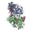
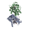
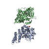
| Title | Crystal Structure of the Potassium Transporter TrkH from Vibrio parahaemolyticus | ||||||
 Components Components | Potassium uptake protein TrkH | ||||||
 Keywords Keywords |  TRANSPORT PROTEIN / structural genomics / PSI-2 / Protein Structure Initiative / New York Consortium on Membrane Protein Structure / NYCOMPS / potassium transport / membrane TRANSPORT PROTEIN / structural genomics / PSI-2 / Protein Structure Initiative / New York Consortium on Membrane Protein Structure / NYCOMPS / potassium transport / membrane | ||||||
| Function / homology |  Function and homology information Function and homology informationpotassium:chloride symporter activity / potassium ion binding / potassium channel activity / potassium ion transmembrane transport / protein homodimerization activity / identical protein binding / plasma membraneSimilarity search - Function | ||||||
| Biological species |   Vibrio parahaemolyticus (bacteria) Vibrio parahaemolyticus (bacteria) | ||||||
| Method | X-RAY DIFFRACTION / SYNCHROTRON / SAD / Resolution: 3.506 Å | ||||||
 Authors Authors | Cao, Y. / Jin, X. / Huang, H. / Levin, E.J. / Zhou, M. / New York Consortium on Membrane Protein Structure (NYCOMPS) | ||||||
 Citation Citation | Journal: Nature / Year: 2011 Title: Crystal structure of a potassium ion transporter, TrkH. Authors: Cao, Y. / Jin, X. / Huang, H. / Derebe, M.G. / Levin, E.J. / Kabaleeswaran, V. / Pan, Y. / Punta, M. / Love, J. / Weng, J. / Quick, M. / Ye, S. / Kloss, B. / Bruni, R. / Martinez-Hackert, E. ...Authors: Cao, Y. / Jin, X. / Huang, H. / Derebe, M.G. / Levin, E.J. / Kabaleeswaran, V. / Pan, Y. / Punta, M. / Love, J. / Weng, J. / Quick, M. / Ye, S. / Kloss, B. / Bruni, R. / Martinez-Hackert, E. / Hendrickson, W.A. / Rost, B. / Javitch, J.A. / Rajashankar, K.R. / Jiang, Y. / Zhou, M. | ||||||
| History |
|
- Structure visualization
| Structure viewer | Molecule: MolmilJmol/JSmol |
|---|
- Downloads & links
-Download
| PDBx/mmCIF format | 3pjz.cif.gz | 185.7 KB | Display | PDBx/mmCIF format |
|---|---|---|---|---|
| PDB format | pdb3pjz.ent.gz | 153.7 KB | Display | PDB format |
| PDBx/mmJSON format | 3pjz.json.gz | Tree view | PDBx/mmJSON format | |
| Others |  Other downloads Other downloads |
-Validation report
| Arichive directory | https://data.pdbj.org/pub/pdb/validation_reports/pj/3pjzftp://data.pdbj.org/pub/pdb/validation_reports/pj/3pjz | HTTPS FTP |
|---|
-Related structure data
| Similar structure data | |
|---|---|
| Other databases |


-Links
 PDBj
PDBj- Assembly
| Deposited unit | 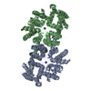
| |||||||||||||||||||||||||||||||||||||||
|---|---|---|---|---|---|---|---|---|---|---|---|---|---|---|---|---|---|---|---|---|---|---|---|---|---|---|---|---|---|---|---|---|---|---|---|---|---|---|---|---|
| 1 |
| |||||||||||||||||||||||||||||||||||||||
| Unit cell |
| |||||||||||||||||||||||||||||||||||||||
| Noncrystallographic symmetry (NCS) | NCS domain:
NCS domain segments:
NCS oper: (Code: given Matrix: (-0.943618, -0.329577, -0.031037), Vector : |
-Components
| #1: Protein | Mass: 54112.457 Da / Num. of mol.: 2 Source method: isolated from a genetically manipulated source Source: (gene. exp.) Vibrio parahaemolyticus (bacteria) / Gene: VP0032 / Plasmid: pET / Production host: Escherichia coli (E. coli) / Strain (production host): BL21(DE3) / References: UniProt: Q87TN7#2: Chemical |   Mass: 39.098 Da / Num. of mol.: 2 / Source method: obtained synthetically / Formula: K Mass: 39.098 Da / Num. of mol.: 2 / Source method: obtained synthetically / Formula: K |
|---|
-Experimental details
-Experiment
| Experiment | Method: X-RAY DIFFRACTION / Number of used crystals: 1 |
|---|
- Sample preparation
| Crystal | Density Matthews: 4.03 Å3/Da / Density % sol: 69.46 % |
|---|---|
| Crystal grow | Temperature: 298 K / Method: microbatch / pH: 5.3 Details: 35% PEG400, 200 mM calcium acetate, 100 mM sodium acetate, pH 5.3, microbatch, temperature 298K |
-Data collection
| Diffraction |
| |||||||||||||||||||||||||||||||||||||||||||||||||||||||
|---|---|---|---|---|---|---|---|---|---|---|---|---|---|---|---|---|---|---|---|---|---|---|---|---|---|---|---|---|---|---|---|---|---|---|---|---|---|---|---|---|---|---|---|---|---|---|---|---|---|---|---|---|---|---|---|---|
| Diffraction source |
| |||||||||||||||||||||||||||||||||||||||||||||||||||||||
| Detector |
| |||||||||||||||||||||||||||||||||||||||||||||||||||||||
| Radiation |
| |||||||||||||||||||||||||||||||||||||||||||||||||||||||
| Radiation wavelength |
| |||||||||||||||||||||||||||||||||||||||||||||||||||||||
| Reflection | Resolution: 3.5→50 Å / Num. obs: 20487 / % possible obs: 89.5 % / Biso Wilson estimate: 63.53 Å2 / Rmerge(I) obs: 0.119 / Χ2: 1.079 / Net I/σ(I): 8.9 | |||||||||||||||||||||||||||||||||||||||||||||||||||||||
| Reflection shell |
|

-Phasing
| Phasing | Method: SAD |
|---|
- Processing
| Software |
| ||||||||||||||||||||||||||||||||||||||||||||||||||||||||
|---|---|---|---|---|---|---|---|---|---|---|---|---|---|---|---|---|---|---|---|---|---|---|---|---|---|---|---|---|---|---|---|---|---|---|---|---|---|---|---|---|---|---|---|---|---|---|---|---|---|---|---|---|---|---|---|---|---|
| Refinement | Method to determine structure: SAD / Resolution: 3.506→14.995 Å / Occupancy max: 1 / Occupancy min: 1 / FOM work R set: 0.7694 / SU ML: 0.54 / σ(F): 1.34 / Phase error: 29.35 / Stereochemistry target values: ML
| ||||||||||||||||||||||||||||||||||||||||||||||||||||||||
| Solvent computation | Shrinkage radii: 0.9 Å / VDW probe radii: 1.11 Å / Solvent model: FLAT BULK SOLVENT MODEL / Bsol: 13.801 Å2 / ksol: 0.229 e/Å3 | ||||||||||||||||||||||||||||||||||||||||||||||||||||||||
| Displacement parameters | Biso max: 195.3 Å2 / Biso mean: 60.4207 Å2 / Biso min: 13.04 Å2
| ||||||||||||||||||||||||||||||||||||||||||||||||||||||||
| Refinement step | Cycle: LAST / Resolution: 3.506→14.995 Å
| ||||||||||||||||||||||||||||||||||||||||||||||||||||||||
| Refine LS restraints |
| ||||||||||||||||||||||||||||||||||||||||||||||||||||||||
| Refine LS restraints NCS |
| ||||||||||||||||||||||||||||||||||||||||||||||||||||||||
| LS refinement shell | Refine-ID: X-RAY DIFFRACTION / Total num. of bins used: 7
|
