[English] 日本語
Yorodumi
- PDB-3gpe: Crystal Structure Analysis of PKC (alpha)-C2 domain complexed wit... -

+
Open data


ID or keywords:

Loading...

-
Basic information

Entry
Database: PDB / ID: 3gpe
TitleCrystal Structure Analysis of PKC (alpha)-C2 domain complexed with Ca2+ and PtdIns(4,5)P2
ComponentsProtein kinase C alpha type
KeywordsSIGNALING PROTEIN / CALCIUM/PHOSPHOLIPID BINDING DOMAIN / C2 domain / Phosphatidil serine / protein kinase / ATP-binding / Kinase / Metal-binding / Nucleotide-binding / Phorbol-ester binding / Phosphoprotein / Serine/threonine-protein kinase / Transferase / Zinc-finger
Function / homology
Function and homology information


Acetylcholine regulates insulin secretion / EGFR Transactivation by Gastrin / HuR (ELAVL1) binds and stabilizes mRNA / Depolymerization of the Nuclear Lamina / Signaling by SCF-KIT / Regulation of KIT signaling / Syndecan interactions / Calmodulin induced events / Disinhibition of SNARE formation / SHC1 events in ERBB2 signaling ...Acetylcholine regulates insulin secretion / EGFR Transactivation by Gastrin / HuR (ELAVL1) binds and stabilizes mRNA / Depolymerization of the Nuclear Lamina / Signaling by SCF-KIT / Regulation of KIT signaling / Syndecan interactions / Calmodulin induced events / Disinhibition of SNARE formation / SHC1 events in ERBB2 signaling / Response to elevated platelet cytosolic Ca2+ / positive regulation of angiotensin-activated signaling pathway / desmosome assembly / positive regulation of dense core granule biogenesis / histone H3T6 kinase activity / cone photoreceptor outer segment / regulation of receptor-mediated endocytosis / VEGFR2 mediated cell proliferation / calcium,diacylglycerol-dependent serine/threonine kinase activity / ooplasm / RET signaling / central nervous system neuron axonogenesis / WNT5A-dependent internalization of FZD4 / RHO GTPases Activate NADPH Oxidases / protein kinase C / cellular response to carbohydrate stimulus / negative regulation of glial cell apoptotic process / diacylglycerol-dependent serine/threonine kinase activity / regulation of platelet aggregation / regulation of muscle contraction / regulation of response to osmotic stress / positive regulation of macrophage differentiation / positive regulation of lipopolysaccharide-mediated signaling pathway / alphav-beta3 integrin-PKCalpha complex / regulation of the force of heart contraction / induction of positive chemotaxis / negative regulation of glucose import / presynaptic cytosol / positive regulation of synapse assembly / regulation of synaptic vesicle exocytosis / muscle cell cellular homeostasis / response to corticosterone / negative regulation of MAPK cascade / positive regulation of cardiac muscle hypertrophy / positive regulation of exocytosis / Trafficking of GluR2-containing AMPA receptors / calyx of Held / positive regulation of cell adhesion / intercalated disc / positive regulation of bone resorption / positive regulation of blood vessel endothelial cell migration / chondrocyte differentiation / response to mechanical stimulus / regulation of peptidyl-tyrosine phosphorylation / negative regulation of insulin receptor signaling pathway / presynaptic modulation of chemical synaptic transmission / positive regulation of endothelial cell proliferation / post-translational protein modification / positive regulation of mitotic cell cycle / response to interleukin-1 / intrinsic apoptotic signaling pathway / neutrophil chemotaxis / positive regulation of endothelial cell migration / response to reactive oxygen species / ciliary basal body / negative regulation of protein phosphorylation / stem cell differentiation / mitochondrial membrane / positive regulation of smooth muscle cell proliferation / peptidyl-threonine phosphorylation / establishment of protein localization / response to organic cyclic compound / response to toxic substance / response to peptide hormone / intracellular calcium ion homeostasis / positive regulation of inflammatory response / positive regulation of angiogenesis / integrin binding / presynapse / response to estradiol / apical part of cell / angiogenesis / response to ethanol / cell population proliferation / negative regulation of translation / learning or memory / positive regulation of ERK1 and ERK2 cascade / cell adhesion / protein kinase activity / intracellular signal transduction / positive regulation of cell migration / positive regulation of protein phosphorylation / axon / negative regulation of cell population proliferation / protein serine kinase activity / protein serine/threonine kinase activity / neuronal cell body / dendrite / lipid binding / perinuclear region of cytoplasm
Similarity search - Function
Classical Protein Kinase C alpha, catalytic domain / Protein kinase C, alpha/beta/gamma types / Protein kinase, C-terminal / Protein kinase C terminal domain / Diacylglycerol/phorbol-ester binding / C2 domain / Phorbol esters/diacylglycerol binding domain (C1 domain) / Zinc finger phorbol-ester/DAG-type signature. / C2 domain / Protein kinase C conserved region 2 (CalB) ...Classical Protein Kinase C alpha, catalytic domain / Protein kinase C, alpha/beta/gamma types / Protein kinase, C-terminal / Protein kinase C terminal domain / Diacylglycerol/phorbol-ester binding / C2 domain / Phorbol esters/diacylglycerol binding domain (C1 domain) / Zinc finger phorbol-ester/DAG-type signature. / C2 domain / Protein kinase C conserved region 2 (CalB) / Zinc finger phorbol-ester/DAG-type profile. / Protein kinase C conserved region 1 (C1) domains (Cysteine-rich domains) / Protein kinase C-like, phorbol ester/diacylglycerol-binding domain / C2 domain / C2 domain profile. / C1-like domain superfamily / Extension to Ser/Thr-type protein kinases / AGC-kinase, C-terminal / AGC-kinase C-terminal domain profile. / C2 domain superfamily / Serine/threonine-protein kinase, active site / Serine/Threonine protein kinases active-site signature. / Protein kinase domain / Serine/Threonine protein kinases, catalytic domain / Protein kinase, ATP binding site / Protein kinases ATP-binding region signature. / Protein kinase domain profile. / Protein kinase domain / Protein kinase-like domain superfamily / Immunoglobulin-like / Sandwich / Mainly Beta
Similarity search - Domain/homology
PHOSPHATE ION / Chem-PT5 / Protein kinase C alpha type
Similarity search - Component
Biological speciesRattus norvegicus (Norway rat)
MethodX-RAY DIFFRACTION / SYNCHROTRON / MOLECULAR REPLACEMENT / Resolution: 2 Å
AuthorsFerrer-Orta, C. / Querol-Audi, J. / Fita, I. / Verdaguer, N.
CitationJournal: Proc.Natl.Acad.Sci.USA / Year: 2009
Title: Structural and mechanistic insights into the association of PKCalpha-C2 domain to PtdIns(4,5)P2.
Authors: Guerrero-Valero, M. / Ferrer-Orta, C. / Querol-Audi, J. / Marin-Vicente, C. / Fita, I. / Gomez-Fernandez, J.C. / Verdaguer, N. / Corbalan-Garcia, S.
History
DepositionMar 23, 2009Deposition site: RCSB / Processing site: RCSB
Revision 1.0May 5, 2009Provider: repository / Type: Initial release
Revision 1.1Jul 13, 2011Group: Advisory / Version format compliance
Revision 1.2Feb 21, 2024Group: Advisory / Data collection ...Advisory / Data collection / Database references / Derived calculations / Structure summary
Category: chem_comp / chem_comp_atom ...chem_comp / chem_comp_atom / chem_comp_bond / database_2 / entity / pdbx_entity_nonpoly / pdbx_unobs_or_zero_occ_atoms / struct_conn / struct_site
Item: _chem_comp.name / _database_2.pdbx_DOI ..._chem_comp.name / _database_2.pdbx_DOI / _database_2.pdbx_database_accession / _entity.pdbx_description / _pdbx_entity_nonpoly.name / _pdbx_unobs_or_zero_occ_atoms.auth_atom_id / _pdbx_unobs_or_zero_occ_atoms.label_atom_id / _struct_conn.pdbx_dist_value / _struct_conn.ptnr1_auth_comp_id / _struct_conn.ptnr1_auth_seq_id / _struct_conn.ptnr1_label_asym_id / _struct_conn.ptnr1_label_atom_id / _struct_conn.ptnr1_label_comp_id / _struct_conn.ptnr1_label_seq_id / _struct_conn.ptnr2_auth_comp_id / _struct_conn.ptnr2_auth_seq_id / _struct_conn.ptnr2_label_asym_id / _struct_conn.ptnr2_label_atom_id / _struct_conn.ptnr2_label_comp_id / _struct_site.pdbx_auth_asym_id / _struct_site.pdbx_auth_comp_id / _struct_site.pdbx_auth_seq_id
Revision 1.3Apr 3, 2024Group: Refinement description / Category: pdbx_initial_refinement_model

-
Structure visualization

Structure viewerMolecule:
MolmilJmol/JSmol

Downloads & links

-
Assembly

Deposited unit
A: Protein kinase C alpha type
hetero molecules


Theoretical massNumber of molelcules
Total (without water)17,3086
Polymers16,0451
Non-polymers1,2625
Water1,65792
1


  • Idetical with deposited unit
  • defined by author&software
TypeNameSymmetry operationNumber
identity operation1_555x,y,z1
MethodPISA
Unit cell
Length a, b, c (Å)57.848, 57.848, 90.479
Angle α, β, γ (deg.)90.00, 90.00, 120.00
Int Tables number154
Space group name H-MP3221

-
Components

#1: Protein Protein kinase C alpha type / PKC-alpha / PKC-A


Mass: 16045.253 Da / Num. of mol.: 1 / Fragment: C2 domain, UNP residues 156-292
Source method: isolated from a genetically manipulated source
Source: (gene. exp.) Rattus norvegicus (Norway rat) / Strain: NORWAY RAT / Gene: Prkca, Pkca / Plasmid: pET28c / Production host: Escherichia coli (E. coli) / Strain (production host): BL21 (DE3) / References: UniProt: P05696, protein kinase C
#2: Chemical ChemComp-CA / CALCIUM ION


Mass: 40.078 Da / Num. of mol.: 3 / Source method: obtained synthetically / Formula: Ca
#3: Chemical ChemComp-PO4 / PHOSPHATE ION / Phosphate


Mass: 94.971 Da / Num. of mol.: 1 / Source method: obtained synthetically / Formula: PO4
#4: Chemical ChemComp-PT5 / [(2R)-1-octadecanoyloxy-3-[oxidanyl-[(1R,2R,3S,4R,5R,6S)-2,3,6-tris(oxidanyl)-4,5-diphosphonooxy-cyclohexyl]oxy-phospho ryl]oxy-propan-2-yl] (8Z)-icosa-5,8,11,14-tetraenoate / Phosphatidylinositol 4,5-bisphosphate / PtdIns(4,5)P2 / Phosphatidylinositol 4,5-bisphosphate


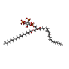
Mass: 1047.088 Da / Num. of mol.: 1 / Source method: obtained synthetically / Formula: C47H85O19P3 / Comment: phospholipid*YM
#5: Water ChemComp-HOH / water / Water


Mass: 18.015 Da / Num. of mol.: 92 / Source method: isolated from a natural source / Formula: H2O

-
Experimental details

-
Experiment

ExperimentMethod: X-RAY DIFFRACTION / Number of used crystals: 1

-
Sample preparation

CrystalDensity Matthews: 2.72 Å3/Da / Density % sol: 54.84 %
Crystal growTemperature: 293 K / Method: vapor diffusion, hanging drop / pH: 6.5
Details: 20% PEG 8000, 50mM potassium phosphate, pH 6.5, VAPOR DIFFUSION, HANGING DROP, temperature 293K

-
Data collection

DiffractionMean temperature: 96 K
Diffraction sourceSource: SYNCHROTRON / Site: ESRF / Beamline: ID14-2 / Wavelength: 0.933
DetectorType: ADSC QUANTUM 4 / Detector: CCD / Date: Sep 1, 2007
RadiationProtocol: SINGLE WAVELENGTH / Monochromatic (M) / Laue (L): M / Scattering type: x-ray
Radiation wavelengthWavelength: 0.933 Å / Relative weight: 1
ReflectionResolution: 1.9→30 Å / Num. obs: 14140 / % possible obs: 99 % / Observed criterion σ(F): 2 / Observed criterion σ(I): 2 / Redundancy: 4 % / Rsym value: 0.06 / Net I/σ(I): 13.3
Reflection shellResolution: 1.9→2 Å / Mean I/σ(I) obs: 1.6 / Rsym value: 0.464 / % possible all: 97.3

-
Processing

Software
NameVersionClassification
ADSCQuantumdata collection
AMoREphasing
REFMAC5.5.0066refinement
MOSFLMdata reduction
SCALAdata scaling
RefinementMethod to determine structure: MOLECULAR REPLACEMENT
Starting model: C2 DOMAIN FROM PROTEIN KINASE C (ALPHA) COMPLEXED WITH CA2+ AND PHOSPHATIDYLSERINE

Resolution: 2→20 Å / Cor.coef. Fo:Fc: 0.905 / Cor.coef. Fo:Fc free: 0.875 / SU B: 9.532 / SU ML: 0.129 / TLS residual ADP flag: LIKELY RESIDUAL / Cross valid method: THROUGHOUT / ESU R: 0.216 / ESU R Free: 0.186 / Stereochemistry target values: MAXIMUM LIKELIHOOD / Details: HYDROGENS HAVE BEEN ADDED IN THE RIDING POSITIONS
RfactorNum. reflection% reflectionSelection details
Rfree0.2773 609 5 %RANDOM
Rwork0.24274 ---
obs0.24434 11566 98.8 %-
Solvent computationIon probe radii: 0.8 Å / Shrinkage radii: 0.8 Å / VDW probe radii: 1.2 Å / Solvent model: MASK
Displacement parametersBiso mean: 16.343 Å2
Baniso -1Baniso -2Baniso -3
1--0.08 Å2-0.04 Å20 Å2
2---0.08 Å20 Å2
3---0.13 Å2
Refinement stepCycle: LAST / Resolution: 2→20 Å
ProteinNucleic acidLigandSolventTotal
Num. atoms1124 0 32 92 1248
Refine LS restraints
Refine-IDTypeDev idealDev ideal targetNumber
X-RAY DIFFRACTIONr_bond_refined_d0.0020.0221180
X-RAY DIFFRACTIONr_angle_refined_deg0.7072.0071603
X-RAY DIFFRACTIONr_dihedral_angle_1_deg4.4235136
X-RAY DIFFRACTIONr_dihedral_angle_2_deg27.33224.3453
X-RAY DIFFRACTIONr_dihedral_angle_3_deg9.42215218
X-RAY DIFFRACTIONr_dihedral_angle_4_deg8.357158
X-RAY DIFFRACTIONr_chiral_restr0.0380.2172
X-RAY DIFFRACTIONr_gen_planes_refined0.0020.021862
X-RAY DIFFRACTIONr_mcbond_it0.1311.5688
X-RAY DIFFRACTIONr_mcangle_it0.25921123
X-RAY DIFFRACTIONr_scbond_it0.3843492
X-RAY DIFFRACTIONr_scangle_it0.6414.5480
LS refinement shellResolution: 2→2.051 Å / Total num. of bins used: 20
RfactorNum. reflection% reflection
Rfree0.296 52 -
Rwork0.271 803 -
obs--98.62 %
Refinement TLS params.Method: refined / Origin x: 26.667 Å / Origin y: -13.927 Å / Origin z: 9.301 Å
111213212223313233
T0.0455 Å20.0358 Å20.0334 Å2-0.0303 Å20.0296 Å2--0.0308 Å2
L5.7297 °2-1.6464 °2-1.8974 °2-4.269 °22.297 °2--5.1862 °2
S0.4083 Å °0.3566 Å °0.3663 Å °-0.2308 Å °-0.1464 Å °-0.0635 Å °-0.3689 Å °-0.3045 Å °-0.2618 Å °

+
About Yorodumi

-
News

-
Feb 9, 2022. New format data for meta-information of EMDB entries

New format data for meta-information of EMDB entries

  • Version 3 of the EMDB header file is now the official format.
  • The previous official version 1.9 will be removed from the archive.

Related info.:EMDB header

External links:wwPDB to switch to version 3 of the EMDB data model

-
Aug 12, 2020. Covid-19 info

Covid-19 info

URL: https://pdbj.org/emnavi/covid19.php

New page: Covid-19 featured information page in EM Navigator.

Related info.:Covid-19 info / Mar 5, 2020. Novel coronavirus structure data

+
Mar 5, 2020. Novel coronavirus structure data

Novel coronavirus structure data

Related info.:Yorodumi Speices / Aug 12, 2020. Covid-19 info

External links:COVID-19 featured content - PDBj / Molecule of the Month (242):Coronavirus Proteases

+
Jan 31, 2019. EMDB accession codes are about to change! (news from PDBe EMDB page)

EMDB accession codes are about to change! (news from PDBe EMDB page)

  • The allocation of 4 digits for EMDB accession codes will soon come to an end. Whilst these codes will remain in use, new EMDB accession codes will include an additional digit and will expand incrementally as the available range of codes is exhausted. The current 4-digit format prefixed with “EMD-” (i.e. EMD-XXXX) will advance to a 5-digit format (i.e. EMD-XXXXX), and so on. It is currently estimated that the 4-digit codes will be depleted around Spring 2019, at which point the 5-digit format will come into force.
  • The EM Navigator/Yorodumi systems omit the EMD- prefix.

Related info.:Q: What is EMD? / ID/Accession-code notation in Yorodumi/EM Navigator

External links:EMDB Accession Codes are Changing Soon! / Contact to PDBj

+
Jul 12, 2017. Major update of PDB

Major update of PDB

  • wwPDB released updated PDB data conforming to the new PDBx/mmCIF dictionary.
  • This is a major update changing the version number from 4 to 5, and with Remediation, in which all the entries are updated.
  • In this update, many items about electron microscopy experimental information are reorganized (e.g. em_software).
  • Now, EM Navigator and Yorodumi are based on the updated data.

External links:wwPDB Remediation / Enriched Model Files Conforming to OneDep Data Standards Now Available in the PDB FTP Archive

-
Yorodumi

Thousand views of thousand structures

  • Yorodumi is a browser for structure data from EMDB, PDB, SASBDB, etc.
  • This page is also the successor to EM Navigator detail page, and also detail information page/front-end page for Omokage search.
  • The word "yorodu" (or yorozu) is an old Japanese word meaning "ten thousand". "mi" (miru) is to see.

Related info.:EMDB / PDB / SASBDB / Comparison of 3 databanks / Yorodumi Search / Aug 31, 2016. New EM Navigator & Yorodumi / Yorodumi Papers / Jmol/JSmol / Function and homology information / Changes in new EM Navigator and Yorodumi

Read more