 Movie
Movie Controller

[English] 日本語
 Yorodumi
- PDB-3etb: Crystal structure of the engineered neutralizing antibody M18 com... -
+ Open data

- Basic information
| Entry | Database: PDB / ID: 3etb | ||||||
|---|---|---|---|---|---|---|---|
| Title | Crystal structure of the engineered neutralizing antibody M18 complexed with anthrax protective antigen domain 4 | ||||||
 Components Components |
| ||||||
 Keywords Keywords |  IMMUNE SYSTEM/TOXIN / single-chain FV / monoclonal antibody / immunoglobulin / toxin / antibody-antigen complex / IMMUNE SYSTEM-TOXIN COMPLEX IMMUNE SYSTEM/TOXIN / single-chain FV / monoclonal antibody / immunoglobulin / toxin / antibody-antigen complex / IMMUNE SYSTEM-TOXIN COMPLEX | ||||||
| Function / homology |  Function and homology information Function and homology informationpositive regulation of apoptotic process in another organism / host cell cytosol / negative regulation of MAPK cascade / Uptake and function of anthrax toxins / host cell endosome membrane / protein homooligomerization / toxin activity / host cell plasma membrane / extracellular region / membrane ...positive regulation of apoptotic process in another organism / host cell cytosol / negative regulation of MAPK cascade / Uptake and function of anthrax toxins / host cell endosome membrane / protein homooligomerization / toxin activity / host cell plasma membrane / extracellular region / membrane / identical protein binding / metal ion bindingSimilarity search - Function | ||||||
| Biological species |  Mus musculus (house mouse) Mus musculus (house mouse) Bacillus anthracis (anthrax bacterium) Bacillus anthracis (anthrax bacterium) | ||||||
| Method | X-RAY DIFFRACTION / MOLECULAR REPLACEMENT / Resolution: 3.8 Å | ||||||
 Authors Authors | Monzingo, A.F. / Leysath, C.E. / Barnett, J. / Iverson, B.L. / Georgiou, G. / Robertus, J.D. | ||||||
 Citation Citation | Journal: J.Mol.Biol. / Year: 2009 Title: Crystal structure of the engineered neutralizing antibody M18 complexed to domain 4 of the anthrax protective antigen. Authors: Leysath, C.E. / Monzingo, A.F. / Maynard, J.A. / Barnett, J. / Georgiou, G. / Iverson, B.L. / Robertus, J.D. | ||||||
| History |
|
- Structure visualization
| Structure viewer | Molecule: MolmilJmol/JSmol |
|---|
- Downloads & links
-Download
| PDBx/mmCIF format | 3etb.cif.gz | 262.6 KB | Display | PDBx/mmCIF format |
|---|---|---|---|---|
| PDB format | pdb3etb.ent.gz | 205.9 KB | Display | PDB format |
| PDBx/mmJSON format | 3etb.json.gz | Tree view | PDBx/mmJSON format | |
| Others |  Other downloads Other downloads |
-Validation report
| Arichive directory | https://data.pdbj.org/pub/pdb/validation_reports/et/3etbftp://data.pdbj.org/pub/pdb/validation_reports/et/3etb | HTTPS FTP |
|---|
-Related structure data
| Related structure data |  3esuSC  3esvC  3et9C 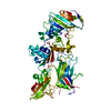 1accS S: Starting model for refinement C: citing same article ( |
|---|---|
| Similar structure data |


-Links
 PDBj
PDBj


- Assembly
| Deposited unit | 
| ||||||||
|---|---|---|---|---|---|---|---|---|---|
| 1 | 
| ||||||||
| 2 | 
| ||||||||
| 3 | 
| ||||||||
| 4 | 
| ||||||||
| Unit cell |
| ||||||||
| Details | Asymmetric unit contains 4 biological units. |
-Components
| #1: Antibody | Mass: 26866.584 Da / Num. of mol.: 4 Mutation: I21V, L46F, S56P, S76N, Q78L, L94P, S1030N, T1058S, K1065E, T1069I Source method: isolated from a genetically manipulated source Details: periplasm / Source: (gene. exp.) Mus musculus (house mouse) / Plasmid: pAK400 / Production host: Escherichia coli (E. coli) / Strain (production host): Jude-1#2: Protein | Mass: 16499.588 Da / Num. of mol.: 4 Fragment: Domain 4 of protective antigen PA-63: UNP residues 621-764 Source method: isolated from a genetically manipulated source Details: secreted / Source: (gene. exp.) Bacillus anthracis (anthrax bacterium) / Gene: pagA, pag, pXO1-110, BXA0164, GBAA_pXO1_0164 / Plasmid: PX01 / Production host: Bacillus anthracis (anthrax bacterium) / Strain (production host): avirulent / References: UniProt: P13423Sequence details | RESIDUE NUMBERS FOR ANTIBODY M18 HEAVY CHAIN FRAGMENT HAVE OFFSET 1000 TO DISTINGUISH THEM FROM THE ...RESIDUE NUMBERS FOR ANTIBODY M18 HEAVY CHAIN FRAGMENT HAVE OFFSET 1000 TO DISTINGUIS | |
|---|
-Experimental details
-Experiment
| Experiment | Method: X-RAY DIFFRACTION / Number of used crystals: 1 |
|---|
- Sample preparation
| Crystal | Density Matthews: 2.91 Å3/Da / Density % sol: 57.68 % |
|---|---|
| Crystal grow | Temperature: 277 K / Method: vapor diffusion, sitting drop / pH: 7.5 Details: 10% PEG 20000, 0.05 M Tris-HCl, pH 7.5, VAPOR DIFFUSION, SITTING DROP, temperature 277K |
-Data collection
| Diffraction | Mean temperature: 100 K |
|---|---|
| Diffraction source | Source: ROTATING ANODE / Type: RIGAKU RUH3R / Wavelength: 1.5418 Å |
| Detector | Type: RIGAKU RAXIS IV++ / Detector: IMAGE PLATE / Date: Jun 27, 2006 / Details: blue max-flux confocal |
| Radiation | Protocol: SINGLE WAVELENGTH / Monochromatic (M) / Laue (L): M / Scattering type: x-ray |
| Radiation wavelength | Wavelength: 1.5418 Å / Relative weight: 1 |
| Reflection | Resolution: 3.8→50 Å / Num. obs: 18357 / % possible obs: 96 % / Observed criterion σ(I): 0 / Redundancy: 2.6 % / Rmerge(I) obs: 0.12 / Χ2: 1.042 / Net I/σ(I): 6.1 |
| Reflection shell | Resolution: 3.8→3.94 Å / Redundancy: 2.2 % / Rmerge(I) obs: 0.283 / Num. unique all: 1705 / Χ2: 1.35 / % possible all: 91.6 |
- Processing
| Software |
| ||||||||||||||||||||||||||||
|---|---|---|---|---|---|---|---|---|---|---|---|---|---|---|---|---|---|---|---|---|---|---|---|---|---|---|---|---|---|
| Refinement | Method to determine structure: MOLECULAR REPLACEMENT Starting model: PDB entries 3ESU, 1ACC Resolution: 3.8→20 Å / Occupancy max: 1 / Occupancy min: 1 / FOM work R set: 0.837 / Isotropic thermal model: isotropic / Cross valid method: THROUGHOUT / σ(F): 0 / Stereochemistry target values: Engh & Huber Details: Non-crystallographic symmetry restraints have been used for positional refinement. Group B factors were refined (2 per residue).
| ||||||||||||||||||||||||||||
| Solvent computation | Bsol: 10 Å2 | ||||||||||||||||||||||||||||
| Displacement parameters | Biso max: 100 Å2 / Biso mean: 13.505 Å2 / Biso min: 10 Å2
| ||||||||||||||||||||||||||||
| Refinement step | Cycle: LAST / Resolution: 3.8→20 Å
| ||||||||||||||||||||||||||||
| Refine LS restraints |
| ||||||||||||||||||||||||||||
| Xplor file | Serial no: 1 / Param file: CNS_TOPPAR:protein_rep.param |
