Biological unit of PpcB is unknown. It appears to be a dimer in the crystal with a buried surface area of 1190 square angstrom calculated with the program SURFACE (CCP4).
-
Components
#1: Protein
Cytochromec3
Mass: 7735.000 Da / Num. of mol.: 2 Source method: isolated from a genetically manipulated source Source: (gene. exp.) Geobacter sulfurreducens (bacteria) / Gene: cyd-1 / Production host: Escherichia coli (E. coli) / Strain (production host): BL21(DE3) / References: UniProt: Q74G83
Resolution: 1.35→50 Å / Num. obs: 58322 / % possible obs: 93 % / Redundancy: 3.3 % / Rmerge(I) obs: 0.089 / Net I/σ(I): 22.4
Reflection shell
Resolution: 1.35→1.4 Å / Redundancy: 1.6 % / Rmerge(I) obs: 0.342 / Mean I/σ(I) obs: 2.5 / Num. unique all: 3851 / % possible all: 62
-
Processing
Software
Name
Version
Classification
REFMAC
5.1.24
refinement
HKL-2000
datareduction
HKL-2000
datascaling
CNS
phasing
Refinement
Method to determine structure: MAD / Resolution: 1.35→19.07 Å / Cor.coef. Fo:Fc: 0.961 / Cor.coef. Fo:Fc free: 0.956 / SU B: 0.931 / SU ML: 0.038 / Cross valid method: THROUGHOUT / ESU R: 0.078 / ESU R Free: 0.063 / Stereochemistry target values: MAXIMUM LIKELIHOOD Details: The number of unique measured reflections is too large because the Bijvoet pairs are treated as separate reflections with the scale anomalous option in HKL2000. The following side chain ...Details: The number of unique measured reflections is too large because the Bijvoet pairs are treated as separate reflections with the scale anomalous option in HKL2000. The following side chain atoms are in weak density and are probably disordered. On Chain A, Asp2: OD1; Lys9: CD, CE, NZ; Lys18: CD, CE, NZ; Lys19: CD, CE, NZ; Lys33: CD, CE, NZ; Lys49: CD, CE, NZ; Lys52: NZ; Glu56: CD, OE1, OE2; Lys60: CD, CE, NZ; Lys70: CG, CD, CE, NZ; Lys71: CB, CG, CD, CE, NZ; on Chain B, Lys9: CG, CD, CE, NZ; Lsy18: NZ; Lys33: CE, NZ; Lys37: CE, NZ; Met58: SD, CE; Lys60: CG, CD, CE, NZ. HYDROGENS HAVE BEEN ADDED IN THE RIDING POSITIONS.
Rfactor
Num. reflection
% reflection
Selection details
Rfree
0.18575
3006
9.9 %
RANDOM
Rwork
0.16168
-
-
-
obs
0.16411
27324
93 %
-
Solvent computation
Ion probe radii: 0.8 Å / Shrinkage radii: 0.8 Å / VDW probe radii: 1.4 Å / Solvent model: BABINET MODEL WITH MASK
Displacement parameters
Biso mean: 7.746 Å2
Baniso -1
Baniso -2
Baniso -3
1-
-0.05 Å2
0 Å2
0 Å2
2-
-
0.06 Å2
0 Å2
3-
-
-
-0.01 Å2
Refinement step
Cycle: LAST / Resolution: 1.35→19.07 Å
Protein
Nucleic acid
Ligand
Solvent
Total
Num. atoms
1076
0
263
201
1540
Refine LS restraints
Refine-ID
Type
Dev ideal
Dev ideal target
Number
X-RAY DIFFRACTION
r_bond_refined_d
0.016
0.022
1406
X-RAY DIFFRACTION
r_bond_other_d
0.002
0.02
1109
X-RAY DIFFRACTION
r_angle_refined_deg
1.648
2.508
1963
X-RAY DIFFRACTION
r_angle_other_deg
1.877
3
2609
X-RAY DIFFRACTION
r_dihedral_angle_1_deg
6.201
5
142
X-RAY DIFFRACTION
r_chiral_restr
0.101
0.2
150
X-RAY DIFFRACTION
r_gen_planes_refined
0.015
0.02
1478
X-RAY DIFFRACTION
r_gen_planes_other
0.029
0.02
190
X-RAY DIFFRACTION
r_nbd_refined
0.404
0.2
410
X-RAY DIFFRACTION
r_nbd_other
0.24
0.2
1368
X-RAY DIFFRACTION
r_nbtor_other
0.092
0.2
648
X-RAY DIFFRACTION
r_xyhbond_nbd_refined
0.161
0.2
140
X-RAY DIFFRACTION
r_metal_ion_refined
0.016
0.2
1
X-RAY DIFFRACTION
r_symmetry_vdw_refined
0.18
0.2
22
X-RAY DIFFRACTION
r_symmetry_vdw_other
0.235
0.2
84
X-RAY DIFFRACTION
r_symmetry_hbond_refined
0.221
0.2
41
X-RAY DIFFRACTION
r_mcbond_it
0.944
1.5
704
X-RAY DIFFRACTION
r_mcangle_it
1.491
2
1123
X-RAY DIFFRACTION
r_scbond_it
1.968
3
702
X-RAY DIFFRACTION
r_scangle_it
2.706
4.5
838
X-RAY DIFFRACTION
r_rigid_bond_restr
1.067
2
1406
X-RAY DIFFRACTION
r_sphericity_free
3.117
2
201
X-RAY DIFFRACTION
r_sphericity_bonded
1.741
2
1341
LS refinement shell
Resolution: 1.35→1.385 Å / Total num. of bins used: 20 /
Rfactor
Num. reflection
Rfree
0.248
124
Rwork
0.219
1286
Refinement TLS params.
Method: refined / Refine-ID: X-RAY DIFFRACTION
ID
L11 (°2)
L12 (°2)
L13 (°2)
L22 (°2)
L23 (°2)
L33 (°2)
S11 (Å °)
S12 (Å °)
S13 (Å °)
S21 (Å °)
S22 (Å °)
S23 (Å °)
S31 (Å °)
S32 (Å °)
S33 (Å °)
T11 (Å2)
T12 (Å2)
T13 (Å2)
T22 (Å2)
T23 (Å2)
T33 (Å2)
Origin x (Å)
Origin y (Å)
Origin z (Å)
1
2.0909
-0.3444
0.026
1.0526
-0.4231
1.09
-0.0176
0.0079
-0.0056
-0.0306
0.0776
-0.0035
0.0108
-0.0715
-0.06
0.0214
0.003
-0.0017
0.0055
-0.0031
0.0098
7.3736
-0.9147
11.8341
2
1.7063
-0.4625
-0.4148
0.6039
-0.018
1.4389
-0.0083
-0.0176
-0.0109
0.0144
0.0321
-0.0138
0.0274
-0.0276
-0.0238
0.023
-0.011
-0.0044
0.006
0.0057
0.0179
-7.7491
-0.6995
-10.1498
Refinement TLS group
ID
Refine-ID
Refine TLS-ID
Auth asym-ID
Label asym-ID
Auth seq-ID
Label seq-ID
1
X-RAY DIFFRACTION
1
A
A - F
1 - 74
1
2
X-RAY DIFFRACTION
2
B
B - I
1 - 74
1
+
About Yorodumi
-
News
-
Feb 9, 2022. New format data for meta-information of EMDB entries
New format data for meta-information of EMDB entries
Version 3 of the EMDB header file is now the official format.
The previous official version 1.9 will be removed from the archive.
In the structure databanks used in Yorodumi, some data are registered as the other names, "COVID-19 virus" and "2019-nCoV". Here are the details of the virus and the list of structure data.
Jan 31, 2019. EMDB accession codes are about to change! (news from PDBe EMDB page)
EMDB accession codes are about to change! (news from PDBe EMDB page)
The allocation of 4 digits for EMDB accession codes will soon come to an end. Whilst these codes will remain in use, new EMDB accession codes will include an additional digit and will expand incrementally as the available range of codes is exhausted. The current 4-digit format prefixed with “EMD-” (i.e. EMD-XXXX) will advance to a 5-digit format (i.e. EMD-XXXXX), and so on. It is currently estimated that the 4-digit codes will be depleted around Spring 2019, at which point the 5-digit format will come into force.
The EM Navigator/Yorodumi systems omit the EMD- prefix.
Related info.:Q: What is EMD? / ID/Accession-code notation in Yorodumi/EM Navigator
Yorodumi is a browser for structure data from EMDB, PDB, SASBDB, etc.
This page is also the successor to EM Navigator detail page, and also detail information page/front-end page for Omokage search.
The word "yorodu" (or yorozu) is an old Japanese word meaning "ten thousand". "mi" (miru) is to see.
Related info.:EMDB / PDB / SASBDB / Comparison of 3 databanks / Yorodumi Search / Aug 31, 2016. New EM Navigator & Yorodumi / Yorodumi Papers / Jmol/JSmol / Function and homology information / Changes in new EM Navigator and Yorodumi
 Movie
Movie Controller


 Open data
 Basic information
 Components
 Keywords
 ELECTRON TRANSPORT / Multiheme cytochromes / cytochrome c7 Geobacter sulfurreducens
 Function and homology information
 Authors
 Citation
 Structure visualization
 Downloads & links
 Other downloads


 PDBj
PDBj
 Assembly

 Mass: 96.063 Da / Num. of mol.: 1 / Source method: obtained synthetically / Formula: SO4
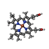
 Mass: 616.487 Da / Num. of mol.: 6 / Source method: obtained synthetically / Formula: C34H32FeN4O4
 Mass: 18.015 Da / Num. of mol.: 201 / Source method: isolated from a natural source / Formula: H2O
 Sample preparation
 / Beamline: 19-BM / Wavelength: 1.73766, 1.73859, 1.78875
 Processing