 Movie
Movie Controller

[English] 日本語
 Yorodumi
- PDB-3bqr: Crystal structure of human death associated protein kinase 3 (DAP... -
+ Open data

- Basic information
| Entry | Database: PDB / ID: 3bqr | ||||||
|---|---|---|---|---|---|---|---|
| Title | Crystal structure of human death associated protein kinase 3 (DAPK3) in complex with an imidazo-pyridazine ligand | ||||||
 Components Components | Death-associated protein kinase 3 | ||||||
 Keywords Keywords |  TRANSFERASE / Death Associated Kinase / DAPK3 / ZIP kinase / ZIPK / DAP kinase 3 / DAP like kinase / Dlk / Structural Genomics Consortium / SGC / Apoptosis / ATP-binding / Chromatin regulator / Nucleotide-binding / Nucleus / Serine/threonine-protein kinase TRANSFERASE / Death Associated Kinase / DAPK3 / ZIP kinase / ZIPK / DAP kinase 3 / DAP like kinase / Dlk / Structural Genomics Consortium / SGC / Apoptosis / ATP-binding / Chromatin regulator / Nucleotide-binding / Nucleus / Serine/threonine-protein kinase | ||||||
| Function / homology |  Function and homology information Function and homology informationregulation of myosin II filament organization / leucine zipper domain binding / cAMP response element binding protein binding / regulation of smooth muscle contraction / regulation of cell motility / Caspase activation via Dependence Receptors in the absence of ligand / regulation of mitotic nuclear division / regulation of focal adhesion assembly / regulation of mitotic cell cycle / regulation of autophagy ...regulation of myosin II filament organization / leucine zipper domain binding / cAMP response element binding protein binding / regulation of smooth muscle contraction / regulation of cell motility / Caspase activation via Dependence Receptors in the absence of ligand / regulation of mitotic nuclear division / regulation of focal adhesion assembly / regulation of mitotic cell cycle / regulation of autophagy / apoptotic signaling pathway / regulation of actin cytoskeleton organization / PML body / small GTPase binding / cellular response to type II interferon / positive regulation of canonical Wnt signaling pathway / chromatin organization / regulation of cell shape / regulation of apoptotic process / protein autophosphorylation / negative regulation of translation / non-specific serine/threonine protein kinase / intracellular signal transduction / positive regulation of cell migration / positive regulation of apoptotic process / protein phosphorylation / protein serine kinase activity / protein serine/threonine kinase activity / apoptotic process / regulation of DNA-templated transcription / protein homodimerization activity / nucleoplasm / ATP binding / identical protein binding / nucleus / cytosol / cytoplasmSimilarity search - Function | ||||||
| Biological species |  Homo sapiens (human) Homo sapiens (human) | ||||||
| Method | X-RAY DIFFRACTION / SYNCHROTRON / MOLECULAR REPLACEMENT / molecular replacement / Resolution: 1.75 Å | ||||||
 Authors Authors | Filippakopoulos, P. / Rellos, P. / Fedorov, O. / Niesen, F. / Pike, A.C.W. / Pilka, E.S. / von Delft, F. / Arrowsmith, C.H. / Edwards, A.M. / Weigelt, J. ...Filippakopoulos, P. / Rellos, P. / Fedorov, O. / Niesen, F. / Pike, A.C.W. / Pilka, E.S. / von Delft, F. / Arrowsmith, C.H. / Edwards, A.M. / Weigelt, J. / Knapp, S. / Structural Genomics Consortium (SGC) | ||||||
 Citation Citation | Journal: To be Published Title: Crystal Structure of Human Death Associated Protein Kinase 3 (DAPK3) in Complex with an Imidazo-Pyridazine Ligand. Authors: Filippakopoulos, P. / Rellos, P. / Fedorov, O. / Niesen, F. / Pike, A.C.W. / Pilka, E.S. / von Delft, F. / Arrowsmith, C.H. / Edwards, A.M. / Weigelt, J. / Knapp, S. | ||||||
| History |
|
- Structure visualization
| Structure viewer | Molecule: MolmilJmol/JSmol |
|---|
- Downloads & links
-Download
| PDBx/mmCIF format | 3bqr.cif.gz | 80.4 KB | Display | PDBx/mmCIF format |
|---|---|---|---|---|
| PDB format | pdb3bqr.ent.gz | 57.2 KB | Display | PDB format |
| PDBx/mmJSON format | 3bqr.json.gz | Tree view | PDBx/mmJSON format | |
| Others |  Other downloads Other downloads |
-Validation report
| Arichive directory | https://data.pdbj.org/pub/pdb/validation_reports/bq/3bqrftp://data.pdbj.org/pub/pdb/validation_reports/bq/3bqr | HTTPS FTP |
|---|
-Related structure data
| Related structure data | 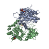 1yrpS S: Starting model for refinement |
|---|---|
| Similar structure data |


-Links
 PDBj
PDBj
- Assembly
| Deposited unit | 
| ||||||||
|---|---|---|---|---|---|---|---|---|---|
| 1 | 
| ||||||||
| Unit cell |
|
-Components
| #1: Protein | Mass: 32545.123 Da / Num. of mol.: 1 / Fragment: Protein kinase domain: Residues 9-289 Source method: isolated from a genetically manipulated source Source: (gene. exp.) Homo sapiens (human) / Gene: DAPK3, ZIPK / Plasmid: pNIC28-Bsa4 / Production host:  Escherichia coli (E. coli) / Strain (production host): BL21(DE3)(R3) Escherichia coli (E. coli) / Strain (production host): BL21(DE3)(R3)References: UniProt: O43293, non-specific serine/threonine protein kinase | ||||
|---|---|---|---|---|---|
| #2: Chemical | ChemComp-4RB /   Mass: 326.350 Da / Num. of mol.: 1 / Source method: obtained synthetically / Formula: C17H18N4O3 Mass: 326.350 Da / Num. of mol.: 1 / Source method: obtained synthetically / Formula: C17H18N4O3 | ||||
| #3: Chemical | Propanediol  Mass: 76.094 Da / Num. of mol.: 2 / Source method: obtained synthetically / Formula: C3H8O2 Mass: 76.094 Da / Num. of mol.: 2 / Source method: obtained synthetically / Formula: C3H8O2#4: Chemical | ChemComp-GOL / | Glycerol  Mass: 92.094 Da / Num. of mol.: 1 / Source method: obtained synthetically / Formula: C3H8O3 Mass: 92.094 Da / Num. of mol.: 1 / Source method: obtained synthetically / Formula: C3H8O3#5: Water | ChemComp-HOH / | Water Mass: 18.015 Da / Num. of mol.: 304 / Source method: isolated from a natural source / Formula: H2O Mass: 18.015 Da / Num. of mol.: 304 / Source method: isolated from a natural source / Formula: H2O |
-Experimental details
-Experiment
| Experiment | Method: X-RAY DIFFRACTION / Number of used crystals: 1 |
|---|
- Sample preparation
| Crystal | Density Matthews: 3.05 Å3/Da / Density % sol: 59.63 % |
|---|---|
| Crystal grow | Temperature: 277 K / Method: vapor diffusion, sitting drop / pH: 6.2 Details: 25% 1,2-propanediol, 10% Glycerol, 0.1M Na/K phosphate pH 6.2, VAPOR DIFFUSION, SITTING DROP, temperature 277K |
-Data collection
| Diffraction | Mean temperature: 100 K | |||||||||||||||||||||||||||||||||||||||||||||||||||||||||||||||||||||||||||||
|---|---|---|---|---|---|---|---|---|---|---|---|---|---|---|---|---|---|---|---|---|---|---|---|---|---|---|---|---|---|---|---|---|---|---|---|---|---|---|---|---|---|---|---|---|---|---|---|---|---|---|---|---|---|---|---|---|---|---|---|---|---|---|---|---|---|---|---|---|---|---|---|---|---|---|---|---|---|---|
| Diffraction source | Source: SYNCHROTRON / Site: SLS  / Beamline: X10SA / Wavelength: 0.98248 Å / Beamline: X10SA / Wavelength: 0.98248 Å | |||||||||||||||||||||||||||||||||||||||||||||||||||||||||||||||||||||||||||||
| Detector | Type: MARMOSAIC 225 mm CCD / Detector: CCD / Date: Oct 26, 2007 | |||||||||||||||||||||||||||||||||||||||||||||||||||||||||||||||||||||||||||||
| Radiation | Monochromator: Si(111) / Protocol: SINGLE WAVELENGTH / Monochromatic (M) / Laue (L): M / Scattering type: x-ray | |||||||||||||||||||||||||||||||||||||||||||||||||||||||||||||||||||||||||||||
| Radiation wavelength | Wavelength: 0.98248 Å / Relative weight: 1 | |||||||||||||||||||||||||||||||||||||||||||||||||||||||||||||||||||||||||||||
| Reflection | Redundancy: 8.5 % / Av σ(I) over netI: 15.1 / Number: 330854 / Rmerge(I) obs: 0.061 / Χ2: 1.02 / D res high: 1.75 Å / D res low: 50 Å / Num. obs: 39150 / % possible obs: 94.8 | |||||||||||||||||||||||||||||||||||||||||||||||||||||||||||||||||||||||||||||
| Diffraction reflection shell |
| |||||||||||||||||||||||||||||||||||||||||||||||||||||||||||||||||||||||||||||
| Reflection | Resolution: 1.75→50 Å / Num. all: 41297 / Num. obs: 39150 / % possible obs: 94.8 % / Redundancy: 8.5 % / Rmerge(I) obs: 0.061 / Rsym value: 0.061 / Χ2: 1.022 / Net I/σ(I): 15.1 | |||||||||||||||||||||||||||||||||||||||||||||||||||||||||||||||||||||||||||||
| Reflection shell | Resolution: 1.75→1.81 Å / Redundancy: 7.4 % / Rmerge(I) obs: 0.894 / Mean I/σ(I) obs: 2.22 / Num. unique all: 3999 / Rsym value: 0.894 / Χ2: 1.025 / % possible all: 98.9 |
-Phasing
| Phasing | Method: molecular replacement | |||||||||
|---|---|---|---|---|---|---|---|---|---|---|
| Phasing MR | Model details: Phaser MODE: MR_AUTO
|
- Processing
| Software |
| |||||||||||||||||||||||||||||||||||||||||||||||||||||||||||||||||||||||||||||||||||||
|---|---|---|---|---|---|---|---|---|---|---|---|---|---|---|---|---|---|---|---|---|---|---|---|---|---|---|---|---|---|---|---|---|---|---|---|---|---|---|---|---|---|---|---|---|---|---|---|---|---|---|---|---|---|---|---|---|---|---|---|---|---|---|---|---|---|---|---|---|---|---|---|---|---|---|---|---|---|---|---|---|---|---|---|---|---|---|
| Refinement | Method to determine structure: MOLECULAR REPLACEMENT Starting model: PDB entry 1YRP Resolution: 1.75→35 Å / Cor.coef. Fo:Fc: 0.965 / Cor.coef. Fo:Fc free: 0.95 / SU B: 5.2 / SU ML: 0.083 / TLS residual ADP flag: LIKELY RESIDUAL / Cross valid method: THROUGHOUT / σ(F): 0 / ESU R: 0.101 / ESU R Free: 0.103 / Stereochemistry target values: MAXIMUM LIKELIHOOD / Details: HYDROGENS HAVE BEEN ADDED IN THE RIDING POSITIONS
| |||||||||||||||||||||||||||||||||||||||||||||||||||||||||||||||||||||||||||||||||||||
| Solvent computation | Ion probe radii: 0.8 Å / Shrinkage radii: 0.8 Å / VDW probe radii: 1.2 Å / Solvent model: MASK | |||||||||||||||||||||||||||||||||||||||||||||||||||||||||||||||||||||||||||||||||||||
| Displacement parameters | Biso mean: 24.304 Å2
| |||||||||||||||||||||||||||||||||||||||||||||||||||||||||||||||||||||||||||||||||||||
| Refinement step | Cycle: LAST / Resolution: 1.75→35 Å
| |||||||||||||||||||||||||||||||||||||||||||||||||||||||||||||||||||||||||||||||||||||
| Refine LS restraints |
| |||||||||||||||||||||||||||||||||||||||||||||||||||||||||||||||||||||||||||||||||||||
| LS refinement shell | Resolution: 1.75→1.795 Å / Total num. of bins used: 20
| |||||||||||||||||||||||||||||||||||||||||||||||||||||||||||||||||||||||||||||||||||||
| Refinement TLS params. | Method: refined / Origin x: 13.7805 Å / Origin y: 42.2295 Å / Origin z: 28.5283 Å
| |||||||||||||||||||||||||||||||||||||||||||||||||||||||||||||||||||||||||||||||||||||
| Refinement TLS group |
|
