 Movie
Movie Controller

[English] 日本語
 Yorodumi
- PDB-3akg: Crystal structure of exo-1,5-alpha-L-arabinofuranosidase complexe... -
+ Open data

- Basic information
| Entry | Database: PDB / ID: 3akg | |||||||||
|---|---|---|---|---|---|---|---|---|---|---|
| Title | Crystal structure of exo-1,5-alpha-L-arabinofuranosidase complexed with alpha-1,5-L-arabinofuranobiose | |||||||||
 Components Components | Putative secreted alpha L-arabinofuranosidase II | |||||||||
 Keywords Keywords |  HYDROLASE / FIVE-BLADED BETA PROPELLER / BETA-TREFOIL HYDROLASE / FIVE-BLADED BETA PROPELLER / BETA-TREFOIL | |||||||||
| Function / homology |  Function and homology information Function and homology informationarabinan catabolic process / L-arabinose metabolic process / non-reducing end alpha-L-arabinofuranosidase / alpha-L-arabinofuranosidase activity / extracellular regionSimilarity search - Function | |||||||||
| Biological species |  Streptomyces avermitilis (bacteria) Streptomyces avermitilis (bacteria) | |||||||||
| Method | X-RAY DIFFRACTION / SYNCHROTRON / MOLECULAR REPLACEMENT / Resolution: 1.8 Å | |||||||||
 Authors Authors | Fujimoto, Z. / Ichinose, H. / Kaneko, S. | |||||||||
 Citation Citation | Journal: J.Biol.Chem. / Year: 2010 Title: Crystal Structure of an Exo-1,5-{alpha}-L-arabinofuranosidase from Streptomyces avermitilis Provides Insights into the Mechanism of Substrate Discrimination between Exo- and Endo-type Enzymes ...Title: Crystal Structure of an Exo-1,5-{alpha}-L-arabinofuranosidase from Streptomyces avermitilis Provides Insights into the Mechanism of Substrate Discrimination between Exo- and Endo-type Enzymes in Glycoside Hydrolase Family 43. Authors: Fujimoto, Z. / Ichinose, H. / Maehara, T. / Honda, M. / Kitaoka, M. / Kaneko, S. #1: Journal: Acta Crystallogr.,Sect.F / Year: 2008 Title: Crystallization and preliminary crystallographic analysis of exo-alpha-1,5-L-arabinofuranosidase from Streptomyces avermitilis NBRC14893. Authors: Fujimoto, Z. / Ichinose, H. / Kaneko, S. #2: Journal: Appl.Microbiol.Biotechnol. / Year: 2008 Title: Characterization of a modular enzyme of exo-1,5-alpha-L-arabinofuranosidase and arabinan binding module from Streptomyces avermitilis NBRC14893. Authors: Ichinose, H. / Yoshida, M. / Fujimoto, Z. / Kaneko, S. | |||||||||
| History |
|
- Structure visualization
| Structure viewer | Molecule: MolmilJmol/JSmol |
|---|
- Downloads & links
-Download
| PDBx/mmCIF format | 3akg.cif.gz | 115.4 KB | Display | PDBx/mmCIF format |
|---|---|---|---|---|
| PDB format | pdb3akg.ent.gz | 84.9 KB | Display | PDB format |
| PDBx/mmJSON format | 3akg.json.gz | Tree view | PDBx/mmJSON format | |
| Others |  Other downloads Other downloads |
-Validation report
| Arichive directory | https://data.pdbj.org/pub/pdb/validation_reports/ak/3akgftp://data.pdbj.org/pub/pdb/validation_reports/ak/3akg | HTTPS FTP |
|---|
-Related structure data
| Related structure data |  3akfSC 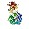 3akhC  3akiC S: Starting model for refinement C: citing same article ( |
|---|---|
| Similar structure data |


-Links
 PDBj
PDBj- Assembly
| Deposited unit | 
| ||||||||
|---|---|---|---|---|---|---|---|---|---|
| 1 |
| ||||||||
| Unit cell |
|
-Components
-Protein , 1 types, 1 molecules A
| #1: Protein | Mass: 51937.340 Da / Num. of mol.: 1 / Fragment: UNP RESIDUES 28-481 Source method: isolated from a genetically manipulated source Source: (gene. exp.) Streptomyces avermitilis (bacteria) / Strain: MA-4680 / Gene: abfA, SAV1043, SAV_1043 / Plasmid: pET30 / Production host: Escherichia coli (E. coli) / Strain (production host): BL21 Gold (DE3)References: UniProt: Q82P90, non-reducing end alpha-L-arabinofuranosidase |
|---|
-Sugars , 2 types, 3 molecules 
| #2: Polysaccharide | alpha-L-arabinofuranose-(1-5)-alpha-L-arabinofuranose / Mass: 282.245 Da / Num. of mol.: 1 Source method: isolated from a genetically manipulated source |
|---|---|
| #5: Sugar | Arabinose Type: L-saccharide, alpha linking / Mass: 150.130 Da / Num. of mol.: 2 Type: L-saccharide, alpha linking / Mass: 150.130 Da / Num. of mol.: 2Source method: isolated from a genetically manipulated source Formula: C5H10O5 |
-Non-polymers , 4 types, 456 molecules 


| #3: Chemical | ChemComp-CL / Chloride Mass: 35.453 Da / Num. of mol.: 1 / Source method: obtained synthetically / Formula: Cl Mass: 35.453 Da / Num. of mol.: 1 / Source method: obtained synthetically / Formula: Cl | ||
|---|---|---|---|
| #4: Chemical | ChemComp-NA /  Mass: 22.990 Da / Num. of mol.: 1 / Source method: obtained synthetically / Formula: Na Mass: 22.990 Da / Num. of mol.: 1 / Source method: obtained synthetically / Formula: Na | ||
| #6: Chemical | Glycerol Mass: 92.094 Da / Num. of mol.: 2 / Source method: obtained synthetically / Formula: C3H8O3 Mass: 92.094 Da / Num. of mol.: 2 / Source method: obtained synthetically / Formula: C3H8O3#7: Water | ChemComp-HOH / | WaterMass: 18.015 Da / Num. of mol.: 452 / Source method: isolated from a natural source / Formula: H2O |
-Experimental details
-Experiment
| Experiment | Method: X-RAY DIFFRACTION / Number of used crystals: 1 |
|---|
- Sample preparation
| Crystal | Density Matthews: 2.51 Å3/Da / Density % sol: 50.94 % |
|---|---|
| Crystal grow | Temperature: 293 K / Method: vapor diffusion, sitting drop / pH: 7 Details: 0.8M sodium citrate, 0.2M sodium chloride, 0.1M Tris, pH 7.0, VAPOR DIFFUSION, SITTING DROP, temperature 293K |
-Data collection
| Diffraction | Mean temperature: 95 K |
|---|---|
| Diffraction source | Source: SYNCHROTRON / Site: Photon Factory  / Beamline: BL-17A / Wavelength: 0.97 Å / Beamline: BL-17A / Wavelength: 0.97 Å |
| Detector | Type: ADSC QUANTUM 270 / Detector: CCD / Date: Jun 26, 2007 |
| Radiation | Monochromator: Numerical link type Si(111) double crystal monochromator Protocol: SINGLE WAVELENGTH / Monochromatic (M) / Laue (L): M / Scattering type: x-ray |
| Radiation wavelength | Wavelength: 0.97 Å / Relative weight: 1 |
| Reflection | Resolution: 1.8→40.42 Å / Num. obs: 47867 / % possible obs: 99.3 % / Observed criterion σ(F): 0 / Observed criterion σ(I): -3 / Redundancy: 13.9 % / Biso Wilson estimate: 25.6 Å2 / Rmerge(I) obs: 0.057 / Rsym value: 0.057 / Net I/σ(I): 52.7 |
| Reflection shell | Resolution: 1.8→1.86 Å / Redundancy: 13.5 % / Rmerge(I) obs: 0.267 / Mean I/σ(I) obs: 9.6 / Num. unique all: 4726 / Rsym value: 0.267 / % possible all: 98.8 |
- Processing
| Software |
| ||||||||||||||||||||||||||||||||||||||||||||||||||||||||||||||||||||||||||||||||||||||||||||||||||||||||||||||||||||||||||||||||||||||||||||||||||||||||||||||||||||||||||
|---|---|---|---|---|---|---|---|---|---|---|---|---|---|---|---|---|---|---|---|---|---|---|---|---|---|---|---|---|---|---|---|---|---|---|---|---|---|---|---|---|---|---|---|---|---|---|---|---|---|---|---|---|---|---|---|---|---|---|---|---|---|---|---|---|---|---|---|---|---|---|---|---|---|---|---|---|---|---|---|---|---|---|---|---|---|---|---|---|---|---|---|---|---|---|---|---|---|---|---|---|---|---|---|---|---|---|---|---|---|---|---|---|---|---|---|---|---|---|---|---|---|---|---|---|---|---|---|---|---|---|---|---|---|---|---|---|---|---|---|---|---|---|---|---|---|---|---|---|---|---|---|---|---|---|---|---|---|---|---|---|---|---|---|---|---|---|---|---|---|---|---|
| Refinement | Method to determine structure: MOLECULAR REPLACEMENT Starting model: PDB ENTRY 3AKF Resolution: 1.8→40.42 Å / Cor.coef. Fo:Fc: 0.958 / Cor.coef. Fo:Fc free: 0.942 / SU B: 2.544 / SU ML: 0.08 / Cross valid method: THROUGHOUT / ESU R Free: 0.121 / Stereochemistry target values: MAXIMUM LIKELIHOOD
| ||||||||||||||||||||||||||||||||||||||||||||||||||||||||||||||||||||||||||||||||||||||||||||||||||||||||||||||||||||||||||||||||||||||||||||||||||||||||||||||||||||||||||
| Solvent computation | Ion probe radii: 0.8 Å / Shrinkage radii: 0.8 Å / VDW probe radii: 1.4 Å / Solvent model: MASK | ||||||||||||||||||||||||||||||||||||||||||||||||||||||||||||||||||||||||||||||||||||||||||||||||||||||||||||||||||||||||||||||||||||||||||||||||||||||||||||||||||||||||||
| Displacement parameters | Biso mean: 25.513 Å2
| ||||||||||||||||||||||||||||||||||||||||||||||||||||||||||||||||||||||||||||||||||||||||||||||||||||||||||||||||||||||||||||||||||||||||||||||||||||||||||||||||||||||||||
| Refine analyze | Luzzati coordinate error obs: 0.0457 Å | ||||||||||||||||||||||||||||||||||||||||||||||||||||||||||||||||||||||||||||||||||||||||||||||||||||||||||||||||||||||||||||||||||||||||||||||||||||||||||||||||||||||||||
| Refinement step | Cycle: LAST / Resolution: 1.8→40.42 Å
| ||||||||||||||||||||||||||||||||||||||||||||||||||||||||||||||||||||||||||||||||||||||||||||||||||||||||||||||||||||||||||||||||||||||||||||||||||||||||||||||||||||||||||
| Refine LS restraints |
| ||||||||||||||||||||||||||||||||||||||||||||||||||||||||||||||||||||||||||||||||||||||||||||||||||||||||||||||||||||||||||||||||||||||||||||||||||||||||||||||||||||||||||
| LS refinement shell | Resolution: 1.802→1.849 Å / Total num. of bins used: 20
|
