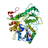
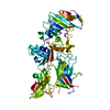
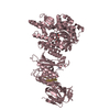
[English] 日本語
Yorodumi
- PDB-2wv9: Crystal Structure of the NS3 protease-helicase from Murray Valley... -

+
Open data


ID or keywords:

Loading...

-
Basic information

Entry
Database: PDB / ID: 2wv9
TitleCrystal Structure of the NS3 protease-helicase from Murray Valley encephalitis virus
ComponentsFLAVIVIRIN PROTEASE NS2B REGULATORY SUBUNIT, FLAVIVIRIN PROTEASE NS3 CATALYTIC SUBUNIT
KeywordsHYDROLASE / NUCLEOTIDE-BINDING / CAPSID PROTEIN / RNA REPLICATION / ENVELOPE PROTEIN / VIRION / HELICASE / FLAVIVIRUS / NUCLEOTIDYLTRANSFERASE
Function / homology
Function and homology information


flavivirin / symbiont-mediated suppression of host JAK-STAT cascade via inhibition of STAT2 activity / symbiont-mediated suppression of host JAK-STAT cascade via inhibition of STAT1 activity / double-stranded RNA binding / viral capsid / nucleoside-triphosphate phosphatase / mRNA (guanine-N7)-methyltransferase / methyltransferase cap1 / clathrin-dependent endocytosis of virus by host cell / mRNA (nucleoside-2'-O-)-methyltransferase activity ...flavivirin / symbiont-mediated suppression of host JAK-STAT cascade via inhibition of STAT2 activity / symbiont-mediated suppression of host JAK-STAT cascade via inhibition of STAT1 activity / double-stranded RNA binding / viral capsid / nucleoside-triphosphate phosphatase / mRNA (guanine-N7)-methyltransferase / methyltransferase cap1 / clathrin-dependent endocytosis of virus by host cell / mRNA (nucleoside-2'-O-)-methyltransferase activity / mRNA 5'-cap (guanine-N7-)-methyltransferase activity / RNA helicase activity / host cell perinuclear region of cytoplasm / host cell endoplasmic reticulum membrane / protein dimerization activity / symbiont-mediated suppression of host type I interferon-mediated signaling pathway / RNA helicase / induction by virus of host autophagy / RNA-directed RNA polymerase / viral RNA genome replication / RNA-dependent RNA polymerase activity / serine-type endopeptidase activity / fusion of virus membrane with host endosome membrane / viral envelope / host cell nucleus / virion attachment to host cell / virion membrane / structural molecule activity / ATP hydrolysis activity / proteolysis / extracellular region / ATP binding / membrane / metal ion binding
Similarity search - Function
Thrombin, subunit H - #120 / : / Flavivirus capsid protein C superfamily / RNA-directed RNA polymerase, thumb domain, Flavivirus / Flavivirus RNA-directed RNA polymerase, thumb domain / Flavivirus envelope glycoprotein E, stem/anchor domain / Flavivirus non-structural protein NS2B / : / Flavivirus NS3 helicase, C-terminal helical domain / Genome polyprotein, Flavivirus ...Thrombin, subunit H - #120 / : / Flavivirus capsid protein C superfamily / RNA-directed RNA polymerase, thumb domain, Flavivirus / Flavivirus RNA-directed RNA polymerase, thumb domain / Flavivirus envelope glycoprotein E, stem/anchor domain / Flavivirus non-structural protein NS2B / : / Flavivirus NS3 helicase, C-terminal helical domain / Genome polyprotein, Flavivirus / Flavivirus non-structural protein NS4A / Flavivirus non-structural protein NS2B / Flavivirus capsid protein C / Flavivirus non-structural protein NS4B / mRNA cap 0/1 methyltransferase / Flavivirus capsid protein C / Flavivirus non-structural protein NS4B / Flavivirus non-structural protein NS4A / Flavivirus NS2B domain profile. / mRNA cap 0 and cap 1 methyltransferase (EC 2.1.1.56 and EC 2.1.1.57) domain profile. / Flavivirus non-structural protein NS2A / Flavivirus non-structural protein NS2A / Flavivirus NS3, petidase S7 / Peptidase S7, Flavivirus NS3 serine protease / Flavivirus NS3 protease (NS3pro) domain profile. / RNA-directed RNA polymerase, flavivirus / Flavivirus RNA-directed RNA polymerase, fingers and palm domains / Flavivirus non-structural Protein NS1 / Flavivirus non-structural protein NS1 / Envelope glycoprotein M, flavivirus / Flavivirus envelope glycoprotein M / Envelope glycoprotein M superfamily, flavivirus / Flavivirus polyprotein propeptide / Flavivirus polyprotein propeptide superfamily / Flavivirus polyprotein propeptide / Flaviviral glycoprotein E, central domain, subdomain 1 / Flaviviral glycoprotein E, central domain, subdomain 2 / Flavivirus envelope glycoprotein E, Stem/Anchor domain / Flavivirus glycoprotein E, immunoglobulin-like domain / Flavivirus envelope glycoprotein E, Stem/Anchor domain superfamily / Flavivirus glycoprotein, immunoglobulin-like domain / Flavivirus glycoprotein central and dimerisation domain / Flavivirus glycoprotein, central and dimerisation domains / Ribosomal RNA methyltransferase, FtsJ domain / FtsJ-like methyltransferase / Flavivirus/Alphavirus glycoprotein, immunoglobulin-like domain superfamily / Flavivirus glycoprotein, central and dimerisation domain superfamily / Flaviviral glycoprotein E, dimerisation domain / DEAD box, Flavivirus / Flavivirus DEAD domain / helicase superfamily c-terminal domain / Immunoglobulin E-set / Superfamilies 1 and 2 helicase C-terminal domain profile. / Superfamilies 1 and 2 helicase ATP-binding type-1 domain profile. / DEAD-like helicases superfamily / Helicase, C-terminal / Helicase superfamily 1/2, ATP-binding domain / Thrombin, subunit H / P-loop containing nucleotide triphosphate hydrolases / RNA-directed RNA polymerase, catalytic domain / RdRp of positive ssRNA viruses catalytic domain profile. / S-adenosyl-L-methionine-dependent methyltransferase superfamily / Peptidase S1, PA clan / DNA/RNA polymerase superfamily / Beta Barrel / P-loop containing nucleoside triphosphate hydrolase / Rossmann fold / 3-Layer(aba) Sandwich / Mainly Beta / Alpha Beta
Similarity search - Domain/homology
Biological speciesMURRAY VALLEY ENCEPHALITIS VIRUS
MethodX-RAY DIFFRACTION / SYNCHROTRON / MOLECULAR REPLACEMENT / Resolution: 2.75 Å
AuthorsAssenberg, R. / Mastrangelo, E. / Walter, T.S. / Verma, A. / Milani, M. / Owens, R.J. / Stuart, D.I. / Grimes, J.M. / Mancini, E.J.
CitationJournal: J.Virol. / Year: 2009
Title: Crystal Structure of a Novel Conformational State of the Flavivirus Ns3 Protein: Implications for Polyprotein Processing and Viral Replication.
Authors: Assenberg, R. / Mastrangelo, E. / Walter, T.S. / Verma, A. / Milani, M. / Owens, R.J. / Stuart, D.I. / Grimes, J.M. / Mancini, E.J.
History
DepositionOct 15, 2009Deposition site: PDBE / Processing site: PDBE
Revision 1.0Dec 1, 2009Provider: repository / Type: Initial release
Revision 1.1Jul 13, 2011Group: Advisory / Version format compliance
Revision 1.2Dec 20, 2023Group: Data collection / Database references ...Data collection / Database references / Other / Refinement description
Category: chem_comp_atom / chem_comp_bond ...chem_comp_atom / chem_comp_bond / database_2 / pdbx_database_status / pdbx_initial_refinement_model
Item: _database_2.pdbx_DOI / _database_2.pdbx_database_accession / _pdbx_database_status.status_code_sf

-
Structure visualization

Structure viewerMolecule:
MolmilJmol/JSmol

Downloads & links

-
Assembly

Deposited unit
A: FLAVIVIRIN PROTEASE NS2B REGULATORY SUBUNIT, FLAVIVIRIN PROTEASE NS3 CATALYTIC SUBUNIT


Theoretical massNumber of molelcules
Total (without water)74,0571
Polymers74,0571
Non-polymers00
Water1,60389
1


  • Idetical with deposited unit
  • defined by author&software
TypeNameSymmetry operationNumber
identity operation1_555x,y,z1
MethodPISA
Unit cell
Length a, b, c (Å)41.910, 105.460, 80.068
Angle α, β, γ (deg.)90.00, 97.42, 90.00
Int Tables number4
Space group name H-MP1211

-
Components

#1: Protein FLAVIVIRIN PROTEASE NS2B REGULATORY SUBUNIT, FLAVIVIRIN PROTEASE NS3 CATALYTIC SUBUNIT


Mass: 74056.555 Da / Num. of mol.: 1 / Fragment: NS2B, RESIDUES 1421-1465
Source method: isolated from a genetically manipulated source
Details: PARTIAL POLYPROTEIN FRAGMENT OF NS2B FRAGMENT SEPARATED FROM NS3 BY A SEQUENCE ENCODING A NINE AMINO ACID LINKER (GGGGSGGGG).
Source: (gene. exp.) MURRAY VALLEY ENCEPHALITIS VIRUS / Strain: MVE-1-51 / Plasmid: OPPF4631 / Production host: ESCHERICHIA COLI (E. coli) / Strain (production host): ROSETTA(DE3)PLYS / References: UniProt: P05769, flavivirin
#2: Water ChemComp-HOH / water / Water


Mass: 18.015 Da / Num. of mol.: 89 / Source method: isolated from a natural source / Formula: H2O
Sequence detailsRESIDUES 49-93 OF PROTEIN NS2B WERE LINKED TO RESIDUES 1-618 OF NS3 VIA A SYNTHETIC LINKER ...RESIDUES 49-93 OF PROTEIN NS2B WERE LINKED TO RESIDUES 1-618 OF NS3 VIA A SYNTHETIC LINKER (GGGGSGGGG) ADDED AT THE C-TERMINUS OF NS2B

-
Experimental details

-
Experiment

ExperimentMethod: X-RAY DIFFRACTION / Number of used crystals: 1

-
Sample preparation

CrystalDensity Matthews: 2.29 Å3/Da / Density % sol: 46.39 % / Description: NONE
Crystal growDetails: 100 MM MES PH 6.0, 20% POLYETHYLENE GLYCOL 6000, 200 MM MAGNESIUM CHLORIDE

-
Data collection

DiffractionMean temperature: 293 K
Diffraction sourceSource: SYNCHROTRON / Site: ESRF / Beamline: ID29 / Wavelength: 1.1271
DetectorType: ADSC CCD / Detector: CCD / Date: May 14, 2007 / Details: MIRRORS
RadiationMonochromator: SI(111) / Protocol: SINGLE WAVELENGTH / Monochromatic (M) / Laue (L): M / Scattering type: x-ray
Radiation wavelengthWavelength: 1.1271 Å / Relative weight: 1
ReflectionResolution: 2.75→43.937 Å / Num. obs: 17750 / % possible obs: 99 % / Observed criterion σ(I): 2 / Redundancy: 7.7 % / Rmerge(I) obs: 0.09 / Net I/σ(I): 19.2
Reflection shellResolution: 2.75→2.82 Å / Redundancy: 7.6 % / Rmerge(I) obs: 0.73 / Mean I/σ(I) obs: 3.2 / % possible all: 98

-
Processing

Software
NameVersionClassification
REFMAC5.5.0102refinement
HKL-2000data reduction
HKL-2000data scaling
PHASERphasing
RefinementMethod to determine structure: MOLECULAR REPLACEMENT
Starting model: PDB ENTRIES 2V8O, 2IJO
Resolution: 2.75→43.94 Å / Cor.coef. Fo:Fc: 0.858 / Cor.coef. Fo:Fc free: 0.844 / SU B: 37.435 / SU ML: 0.353 / TLS residual ADP flag: LIKELY RESIDUAL / Cross valid method: THROUGHOUT / ESU R Free: 0.448 / Stereochemistry target values: MAXIMUM LIKELIHOOD / Details: HYDROGENS HAVE BEEN ADDED IN THE RIDING POSITIONS
RfactorNum. reflection% reflectionSelection details
Rfree0.3 884 5 %RANDOM
Rwork0.267 ---
obs0.268 16866 98.6 %-
Solvent computationIon probe radii: 0.8 Å / Shrinkage radii: 0.8 Å / VDW probe radii: 1.4 Å / Solvent model: BABINET MODEL WITH MASK
Displacement parametersBiso mean: 6.17 Å2
Baniso -1Baniso -2Baniso -3
1--0.29 Å20 Å2-0.24 Å2
2--2.68 Å20 Å2
3----2.46 Å2
Refinement stepCycle: LAST / Resolution: 2.75→43.94 Å
ProteinNucleic acidLigandSolventTotal
Num. atoms4677 0 0 89 4766
Refine LS restraints
Refine-IDTypeDev idealDev ideal targetNumber
X-RAY DIFFRACTIONr_bond_refined_d0.0060.0224785
X-RAY DIFFRACTIONr_bond_other_d
X-RAY DIFFRACTIONr_angle_refined_deg0.8991.9466496
X-RAY DIFFRACTIONr_angle_other_deg
X-RAY DIFFRACTIONr_dihedral_angle_1_deg4.4315596
X-RAY DIFFRACTIONr_dihedral_angle_2_deg30.94523.349215
X-RAY DIFFRACTIONr_dihedral_angle_3_deg13.85415783
X-RAY DIFFRACTIONr_dihedral_angle_4_deg13.5161539
X-RAY DIFFRACTIONr_chiral_restr0.0590.2708
X-RAY DIFFRACTIONr_gen_planes_refined0.0030.0213659
X-RAY DIFFRACTIONr_gen_planes_other
X-RAY DIFFRACTIONr_nbd_refined0.1690.21975
X-RAY DIFFRACTIONr_nbd_other
X-RAY DIFFRACTIONr_nbtor_refined0.2940.23158
X-RAY DIFFRACTIONr_nbtor_other
X-RAY DIFFRACTIONr_xyhbond_nbd_refined0.1120.2153
X-RAY DIFFRACTIONr_xyhbond_nbd_other
X-RAY DIFFRACTIONr_metal_ion_refined
X-RAY DIFFRACTIONr_metal_ion_other
X-RAY DIFFRACTIONr_symmetry_vdw_refined0.1730.256
X-RAY DIFFRACTIONr_symmetry_vdw_other
X-RAY DIFFRACTIONr_symmetry_hbond_refined0.0840.25
X-RAY DIFFRACTIONr_symmetry_hbond_other
X-RAY DIFFRACTIONr_symmetry_metal_ion_refined
X-RAY DIFFRACTIONr_symmetry_metal_ion_other
X-RAY DIFFRACTIONr_mcbond_it0.1451.52977
X-RAY DIFFRACTIONr_mcbond_other
X-RAY DIFFRACTIONr_mcangle_it0.26724792
X-RAY DIFFRACTIONr_mcangle_other
X-RAY DIFFRACTIONr_scbond_it0.24631808
X-RAY DIFFRACTIONr_scbond_other
X-RAY DIFFRACTIONr_scangle_it0.4344.51704
X-RAY DIFFRACTIONr_scangle_other
X-RAY DIFFRACTIONr_long_range_B_refined
X-RAY DIFFRACTIONr_long_range_B_other
X-RAY DIFFRACTIONr_rigid_bond_restr
X-RAY DIFFRACTIONr_sphericity_free
X-RAY DIFFRACTIONr_sphericity_bonded
LS refinement shellResolution: 2.75→2.82 Å / Total num. of bins used: 20
RfactorNum. reflection% reflection
Rfree0.311 66 -
Rwork0.324 1226 -
obs--98.1 %
Refinement TLS params.

Method: refined / Refine-ID: X-RAY DIFFRACTION

IDL112)L122)L132)L222)L232)L332)S11 (Å °)S12 (Å °)S13 (Å °)S21 (Å °)S22 (Å °)S23 (Å °)S31 (Å °)S32 (Å °)S33 (Å °)T112)T122)T132)T222)T232)T332)Origin x (Å)Origin y (Å)Origin z (Å)
16.2672-1.88110.92121.99742.33199.5093-0.01370.0395-0.52250.27870.11160.16060.7028-0.1374-0.0980.29740.0630.0130.3084-0.04790.383627.45718.745-3.284
21.122-0.3592-0.32781.1232-0.78611.662-0.027-0.2287-0.2115-0.1027-0.05660.18190.3450.05280.08370.5660.0454-0.00940.5197-0.06030.4979-1.081.8436.051
338.01680.48492.25134.51756.468622.9086-2.05262.8624.9487-0.13390.4021-0.1866-2.9047-0.24621.65050.7643-0.0214-0.50650.32060.21310.930526.34728.893-8.085
Refinement TLS group
IDRefine-IDRefine TLS-IDAuth asym-IDAuth seq-ID
1X-RAY DIFFRACTION1A18 - 168
2X-RAY DIFFRACTION2A178 - 618
3X-RAY DIFFRACTION3A-52 - -33

+
About Yorodumi

-
News

-
Feb 9, 2022. New format data for meta-information of EMDB entries

New format data for meta-information of EMDB entries

  • Version 3 of the EMDB header file is now the official format.
  • The previous official version 1.9 will be removed from the archive.

Related info.:EMDB header

External links:wwPDB to switch to version 3 of the EMDB data model

-
Aug 12, 2020. Covid-19 info

Covid-19 info

URL: https://pdbj.org/emnavi/covid19.php

New page: Covid-19 featured information page in EM Navigator.

Related info.:Covid-19 info / Mar 5, 2020. Novel coronavirus structure data

+
Mar 5, 2020. Novel coronavirus structure data

Novel coronavirus structure data

Related info.:Yorodumi Speices / Aug 12, 2020. Covid-19 info

External links:COVID-19 featured content - PDBj / Molecule of the Month (242):Coronavirus Proteases

+
Jan 31, 2019. EMDB accession codes are about to change! (news from PDBe EMDB page)

EMDB accession codes are about to change! (news from PDBe EMDB page)

  • The allocation of 4 digits for EMDB accession codes will soon come to an end. Whilst these codes will remain in use, new EMDB accession codes will include an additional digit and will expand incrementally as the available range of codes is exhausted. The current 4-digit format prefixed with “EMD-” (i.e. EMD-XXXX) will advance to a 5-digit format (i.e. EMD-XXXXX), and so on. It is currently estimated that the 4-digit codes will be depleted around Spring 2019, at which point the 5-digit format will come into force.
  • The EM Navigator/Yorodumi systems omit the EMD- prefix.

Related info.:Q: What is EMD? / ID/Accession-code notation in Yorodumi/EM Navigator

External links:EMDB Accession Codes are Changing Soon! / Contact to PDBj

+
Jul 12, 2017. Major update of PDB

Major update of PDB

  • wwPDB released updated PDB data conforming to the new PDBx/mmCIF dictionary.
  • This is a major update changing the version number from 4 to 5, and with Remediation, in which all the entries are updated.
  • In this update, many items about electron microscopy experimental information are reorganized (e.g. em_software).
  • Now, EM Navigator and Yorodumi are based on the updated data.

External links:wwPDB Remediation / Enriched Model Files Conforming to OneDep Data Standards Now Available in the PDB FTP Archive

-
Yorodumi

Thousand views of thousand structures

  • Yorodumi is a browser for structure data from EMDB, PDB, SASBDB, etc.
  • This page is also the successor to EM Navigator detail page, and also detail information page/front-end page for Omokage search.
  • The word "yorodu" (or yorozu) is an old Japanese word meaning "ten thousand". "mi" (miru) is to see.

Related info.:EMDB / PDB / SASBDB / Comparison of 3 databanks / Yorodumi Search / Aug 31, 2016. New EM Navigator & Yorodumi / Yorodumi Papers / Jmol/JSmol / Function and homology information / Changes in new EM Navigator and Yorodumi

Read more