 Movie
Movie Controller


+ Open data

- Basic information
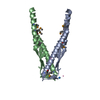
| Entry | Database: PDB / ID: 2wj5 | ||||||
|---|---|---|---|---|---|---|---|
| Title | Rat alpha crystallin domain | ||||||
 Components Components | HEAT SHOCK PROTEIN BETA-6 Heat shock response Heat shock response | ||||||
 Keywords Keywords | CHAPERONE / DISULFIDE BOND / STRESS RESPONSE | ||||||
| Function / homology |  Function and homology informationregulation of muscle contraction / structural constituent of eye lens / negative regulation of cardiac muscle cell apoptotic process / chaperone-mediated protein folding / positive regulation of angiogenesis / unfolded protein binding / protein-folding chaperone binding / nucleolus / Golgi apparatus / protein homodimerization activity ...regulation of muscle contraction / structural constituent of eye lens / negative regulation of cardiac muscle cell apoptotic process / chaperone-mediated protein folding / positive regulation of angiogenesis / unfolded protein binding / protein-folding chaperone binding / nucleolus / Golgi apparatus / protein homodimerization activity / extracellular region / nucleus / cytosol / cytoplasm Function and homology informationregulation of muscle contraction / structural constituent of eye lens / negative regulation of cardiac muscle cell apoptotic process / chaperone-mediated protein folding / positive regulation of angiogenesis / unfolded protein binding / protein-folding chaperone binding / nucleolus / Golgi apparatus / protein homodimerization activity ...regulation of muscle contraction / structural constituent of eye lens / negative regulation of cardiac muscle cell apoptotic process / chaperone-mediated protein folding / positive regulation of angiogenesis / unfolded protein binding / protein-folding chaperone binding / nucleolus / Golgi apparatus / protein homodimerization activity / extracellular region / nucleus / cytosol / cytoplasmSimilarity search - Function | ||||||
| Biological species |  RATTUS NORVEGICUS (Norway rat) RATTUS NORVEGICUS (Norway rat) | ||||||
| Method | X-RAY DIFFRACTION / SYNCHROTRON / MOLECULAR REPLACEMENT / Resolution: 1.12 Å | ||||||
 Authors Authors | Naylor, C.E. / Bagneris, C. / Bateman, O.A. / Cronin, N. / Keep, N.H. / Slingsby, C. | ||||||
 Citation Citation | Journal: J.Mol.Biol. / Year: 2009 Title: Crystal Structures of Alpha-Crystallin Domain Dimers of Alphab-Crystallin and Hsp20. Authors: Bagneris, C. / Bateman, O.A. / Naylor, C.E. / Cronin, N. / Boelens, W.C. / Keep, N.H. / Slingsby, C. | ||||||
| History |
|
- Structure visualization
| Structure viewer | Molecule: MolmilJmol/JSmol |
|---|
- Downloads & links
-Download
| PDBx/mmCIF format | 2wj5.cif.gz | 64.5 KB | Display | PDBx/mmCIF format |
|---|---|---|---|---|
| PDB format | pdb2wj5.ent.gz | 48.4 KB | Display | PDB format |
| PDBx/mmJSON format | 2wj5.json.gz | Tree view | PDBx/mmJSON format | |
| Others |  Other downloads Other downloads |
-Validation report
| Arichive directory | https://data.pdbj.org/pub/pdb/validation_reports/wj/2wj5ftp://data.pdbj.org/pub/pdb/validation_reports/wj/2wj5 | HTTPS FTP |
|---|
-Related structure data
| Related structure data | 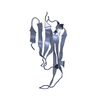 2wj7C  1gmeS C: citing same article ( S: Starting model for refinement |
|---|---|
| Similar structure data |


-Links
 PDBj
PDBj


- Assembly
| Deposited unit | 
| ||||||||
|---|---|---|---|---|---|---|---|---|---|
| 1 |
| ||||||||
| Unit cell |
| ||||||||
| Components on special symmetry positions |
|
-Components
| #1: Protein | Heat shock response / HEAT SHOCK 20 KDA-LIKE PROTEIN P20 / HSPB6 Mass: 10844.139 Da / Num. of mol.: 1 / Fragment: ALPHA-CRYSTALLIN DOMAIN, RESIDUES 65-162 Source method: isolated from a genetically manipulated source Source: (gene. exp.) RATTUS NORVEGICUS (Norway rat) / Production host:  ESCHERICHIA COLI (E. coli) / References: UniProt: P97541 ESCHERICHIA COLI (E. coli) / References: UniProt: P97541 |
|---|---|
| #2: Water | ChemComp-HOH / Water Mass: 18.015 Da / Num. of mol.: 227 / Source method: isolated from a natural source / Formula: H2O Mass: 18.015 Da / Num. of mol.: 227 / Source method: isolated from a natural source / Formula: H2O |
| Sequence details | THE ALPHA-CRYSTALLIN |
-Experimental details
-Experiment
| Experiment | Method: X-RAY DIFFRACTION / Number of used crystals: 1 |
|---|
- Sample preparation
| Crystal | Density Matthews: 2.66 Å3/Da / Density % sol: 54 % / Description: NONE |
|---|---|
| Crystal grow | Temperature: 289 K / Method: vapor diffusion / pH: 6.5 Details: SINGLE CRYSTALS OF RAT HSP20 ACD GREW AT 16 DEGREESIN 100 MM MES PH 6.5, BETWEEN 45-52% V/V PEG 200, AT A PROTEIN CONCENTRATION OF AROUND 10 MG/ML USING 1 UL PROTEIN SOLUTION AND 2 UL RESERVOIR SOLUTION. |
-Data collection
| Diffraction | Mean temperature: 100 K |
|---|---|
| Diffraction source | Source: SYNCHROTRON / Site: Diamond  / Beamline: I04 / Wavelength: 0.9757 / Beamline: I04 / Wavelength: 0.9757 |
| Detector | Type: ADSC CCD / Detector: CCD / Date: Jan 5, 2008 |
| Radiation | Monochromator: SI CRYSTAL / Protocol: SINGLE WAVELENGTH / Monochromatic (M) / Laue (L): M / Scattering type: x-ray |
| Radiation wavelength | Wavelength: 0.9757 Å / Relative weight: 1 |
| Reflection | Resolution: 1.12→28 Å / Num. obs: 39225 / % possible obs: 91.6 % / Observed criterion σ(I): 0 / Redundancy: 6.7 % / Biso Wilson estimate: 8.4 Å2 / Rmerge(I) obs: 0.09 / Net I/σ(I): 10.7 |
| Reflection shell | Resolution: 1.12→1.15 Å / Redundancy: 4.5 % / Rmerge(I) obs: 0.4 / Mean I/σ(I) obs: 2.5 / % possible all: 51.5 |
- Processing
| Software |
| |||||||||||||||||||||||||||||||||||||||||||||||||||||||||||||||||||||||||||||||||||||||||||||||||||||||||
|---|---|---|---|---|---|---|---|---|---|---|---|---|---|---|---|---|---|---|---|---|---|---|---|---|---|---|---|---|---|---|---|---|---|---|---|---|---|---|---|---|---|---|---|---|---|---|---|---|---|---|---|---|---|---|---|---|---|---|---|---|---|---|---|---|---|---|---|---|---|---|---|---|---|---|---|---|---|---|---|---|---|---|---|---|---|---|---|---|---|---|---|---|---|---|---|---|---|---|---|---|---|---|---|---|---|---|
| Refinement | Method to determine structure: MOLECULAR REPLACEMENT Starting model: PDB ENTRY 1GME Resolution: 1.12→28.071 Å / SU ML: 0.09 / σ(F): 1.38 / Phase error: 16.21 / Stereochemistry target values: ML
| |||||||||||||||||||||||||||||||||||||||||||||||||||||||||||||||||||||||||||||||||||||||||||||||||||||||||
| Solvent computation | Shrinkage radii: 0.9 Å / VDW probe radii: 1.11 Å / Solvent model: FLAT BULK SOLVENT MODEL / Bsol: 99 Å2 / ksol: 0.426 e/Å3 | |||||||||||||||||||||||||||||||||||||||||||||||||||||||||||||||||||||||||||||||||||||||||||||||||||||||||
| Displacement parameters | Biso mean: 18.6 Å2
| |||||||||||||||||||||||||||||||||||||||||||||||||||||||||||||||||||||||||||||||||||||||||||||||||||||||||
| Refinement step | Cycle: LAST / Resolution: 1.12→28.071 Å
| |||||||||||||||||||||||||||||||||||||||||||||||||||||||||||||||||||||||||||||||||||||||||||||||||||||||||
| Refine LS restraints |
| |||||||||||||||||||||||||||||||||||||||||||||||||||||||||||||||||||||||||||||||||||||||||||||||||||||||||
| LS refinement shell |
|
