Mass: 18.015 Da / Num. of mol.: 949 / Source method: isolated from a natural source / Formula: H2O
Compound details
ENGINEERED RESIDUE IN CHAIN A, LEU 461 TO ALA ENGINEERED RESIDUE IN CHAIN B, LEU 461 TO ALA ...ENGINEERED RESIDUE IN CHAIN A, LEU 461 TO ALA ENGINEERED RESIDUE IN CHAIN B, LEU 461 TO ALA ENGINEERED RESIDUE IN CHAIN C, LEU 461 TO ALA ENGINEERED RESIDUE IN CHAIN D, LEU 461 TO ALA
Sequence details
RESIDUE 461 HAS BEEN POINT MUTATED FROM LEUCINE INTO ALANINE
-
Experimental details
-
Experiment
Experiment
Method: X-RAY DIFFRACTION / Number of used crystals: 1
-
Sample preparation
Crystal
Density Matthews: 2.32 Å3/Da / Density % sol: 47 % / Description: NONE
-
Data collection
Diffraction
Mean temperature: 100 K
Diffraction source
Source: SYNCHROTRON / Site: MAX II / Beamline: I911-3 / Wavelength: 1
Detector
Type: MARRESEARCH / Detector: CCD / Date: Oct 20, 2006 / Details: RH-COATED SI MIRROR, RH- COATED TOROIDAL SI MIRROR
Radiation
Monochromator: DOUBLE CRYSTAL MONOCHROMATOR, SI(111). / Protocol: SINGLE WAVELENGTH / Monochromatic (M) / Laue (L): M / Scattering type: x-ray
Radiation wavelength
Wavelength: 1 Å / Relative weight: 1
Reflection
Resolution: 2.2→107.8 Å / Num. obs: 111569 / % possible obs: 97 % / Observed criterion σ(I): 6 / Redundancy: 2.9 % / Biso Wilson estimate: 27.8 Å2 / Rmerge(I) obs: 0.12 / Net I/σ(I): 8.8
Reflection shell
Resolution: 2.2→2.32 Å / Redundancy: 2.8 % / Rmerge(I) obs: 0.46 / Mean I/σ(I) obs: 2.2 / % possible all: 99.2
Resolution: 2.2→35 Å / Cor.coef. Fo:Fc: 0.951 / Cor.coef. Fo:Fc free: 0.922 / SU B: 5.879 / SU ML: 0.148 / Cross valid method: THROUGHOUT / ESU R: 0.271 / ESU R Free: 0.207 / Stereochemistry target values: MAXIMUM LIKELIHOOD / Details: HYDROGENS HAVE BEEN ADDED IN THE RIDING POSITIONS.
Rfactor
Num. reflection
% reflection
Selection details
Rfree
0.228
5610
5 %
RANDOM
Rwork
0.18
-
-
-
obs
0.182
105886
96.5 %
-
Solvent computation
Ion probe radii: 0.8 Å / Shrinkage radii: 0.8 Å / VDW probe radii: 1.4 Å / Solvent model: MASK
 Movie
Movie Controller

 Yorodumi
 Open data
 Basic information
 Components
 Keywords
 Function and homology information
 Authors
 Citation
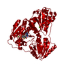
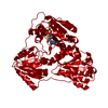
 Structure visualization
 Downloads & links
 Other downloads


 PDBj
PDBj


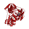
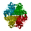
 Assembly

 Mass: 24.305 Da / Num. of mol.: 6 / Source method: obtained synthetically / Formula: Mg
 Mass: 96.063 Da / Num. of mol.: 4 / Source method: obtained synthetically / Formula: SO4
 Mass: 425.314 Da / Num. of mol.: 4 / Source method: obtained synthetically / Formula: C12H19N4O7P2S
 Mass: 18.015 Da / Num. of mol.: 949 / Source method: isolated from a natural source / Formula: H2O
 Sample preparation
 / Beamline: I911-3 / Wavelength: 1
  Processing