BIOMOLECULE: 1 SEE REMARK 350 FOR THE AUTHOR PROVIDED AND PROGRAM GENERATED ASSEMBLY INFORMATION ... BIOMOLECULE: 1 SEE REMARK 350 FOR THE AUTHOR PROVIDED AND PROGRAM GENERATED ASSEMBLY INFORMATION FOR THE STRUCTURE IN THIS ENTRY. THE REMARK MAY ALSO PROVIDE INFORMATION ON BURIED SURFACE AREA. SIZE EXCLUSION CHROMATOGRAPHY SUPPORTS THE ASSIGNMENT OF A DIMER AS THE SIGNIFICANT OLIGOMERIZATION STATE.
Remark 999
SEQUENCE THE CONSTRUCT WAS EXPRESSED WITH A PURIFICATION TAG MGSDKIHHHHHHENLYFQG. THE TAG WAS ... SEQUENCE THE CONSTRUCT WAS EXPRESSED WITH A PURIFICATION TAG MGSDKIHHHHHHENLYFQG. THE TAG WAS REMOVED WITH TEV PROTEASE LEAVING ONLY A GLYCINE FOLLOWED BY THE TARGET SEQUENCE. THE ALTERNATE START CODON HAS BEEN CHANGED TO AUG DURING CLONING TO MAINTAIN A MET AMINO ACID AT POSITION 1.
Type: MARMOSAIC 325 mm CCD / Detector: CCD / Date: Jun 3, 2007 / Details: Flat mirror (vertical focusing)
Radiation
Monochromator: Single crystal Si(111) bent (horizontal focusing) Protocol: MAD / Monochromatic (M) / Laue (L): M / Scattering type: x-ray
Radiation wavelength
ID
Wavelength (Å)
Relative weight
1
0.91837
1
2
0.97901
1
3
0.97926
1
Reflection
Resolution: 1.6→27.995 Å / Num. obs: 101945 / % possible obs: 98.7 % / Observed criterion σ(I): -3 / Redundancy: 3.28 % / Biso Wilson estimate: 18.5 Å2 / Rmerge(I) obs: 0.045 / Net I/σ(I): 9.92
Reflection shell
Resolution (Å)
Rmerge(I) obs
Mean I/σ(I) obs
Num. measured obs
Diffraction-ID
% possible all
1.6-1.66
0.314
2.2
28804
1
94.5
1.66-1.72
0.261
2.8
30168
1
99.5
1.72-1.8
0.213
3.4
34334
1
99.5
1.8-1.9
0.165
4.5
35319
1
99.4
1.9-2.02
0.112
6.6
33788
1
99.2
2.02-2.17
0.076
9.4
32642
1
99.5
2.17-2.39
0.058
12.2
34446
1
99.4
2.39-2.73
0.045
15.1
34248
1
99.3
2.73-3.44
0.034
19
34856
1
99
3.44-27.995
0.025
23.9
35411
1
97.8
-
Phasing
Phasing
Method: MAD
-
Processing
Software
Name
Version
Classification
NB
REFMAC
5.2.0019
refinement
PHENIX
refinement
SOLVE
phasing
MolProbity
3beta29
modelbuilding
XSCALE
datascaling
PDB_EXTRACT
3
dataextraction
MAR345
CCD
datacollection
XDS
datareduction
Refinement
Method to determine structure: MAD / Resolution: 1.6→27.995 Å / Cor.coef. Fo:Fc: 0.97 / Cor.coef. Fo:Fc free: 0.96 / SU B: 2.979 / SU ML: 0.052 / TLS residual ADP flag: LIKELY RESIDUAL / Cross valid method: THROUGHOUT / σ(F): 0 / ESU R: 0.078 / ESU R Free: 0.078 Stereochemistry target values: MAXIMUM LIKELIHOOD WITH PHASES Details: 1. HYDROGENS HAVE BEEN ADDED IN THE RIDING POSITIONS. 2. A MET-INHIBITION PROTOCOL WAS USED FOR SELENOMETHIONINE INCORPORATION DURING PROTEIN EXPRESSION. THE OCCUPANCY OF THE SE ATOMS IN THE ...Details: 1. HYDROGENS HAVE BEEN ADDED IN THE RIDING POSITIONS. 2. A MET-INHIBITION PROTOCOL WAS USED FOR SELENOMETHIONINE INCORPORATION DURING PROTEIN EXPRESSION. THE OCCUPANCY OF THE SE ATOMS IN THE MSE RESIDUES WAS REDUCED TO 0.75 TO ACCOUNT FOR THE REDUCED SCATTERING POWER DUE TO PARTIAL S-MET INCORPORATION. 3. ATOM RECORD CONTAINS RESIDUAL B FACTORS ONLY. 4. RESIDUES 360-363 IN CHAINS A,B ARE DISORDERED AND HAVE NOT BEEN MODELLED. 5. FOUR PEG, ONE PGE AND ONE PG4 MOLECULE(S) HAVE BEEN MODELED AS PART OF THE SOLVENT STRUCTURE.
Rfactor
Num. reflection
% reflection
Selection details
Rfree
0.18
5090
5 %
RANDOM
Rwork
0.154
-
-
-
obs
0.155
101918
99.4 %
-
Solvent computation
Ion probe radii: 0.8 Å / Shrinkage radii: 0.8 Å / VDW probe radii: 1.2 Å / Solvent model: MASK
Displacement parameters
Biso mean: 14.803 Å2
Baniso -1
Baniso -2
Baniso -3
1-
0.46 Å2
0 Å2
-0.75 Å2
2-
-
0.17 Å2
0 Å2
3-
-
-
-0.67 Å2
Refinement step
Cycle: LAST / Resolution: 1.6→27.995 Å
Protein
Nucleic acid
Ligand
Solvent
Total
Num. atoms
5505
0
51
916
6472
Refine LS restraints
Refine-ID
Type
Dev ideal
Dev ideal target
Number
X-RAY DIFFRACTION
r_bond_refined_d
0.017
0.022
5985
X-RAY DIFFRACTION
r_bond_other_d
0.002
0.02
4151
X-RAY DIFFRACTION
r_angle_refined_deg
1.624
1.969
8146
X-RAY DIFFRACTION
r_angle_other_deg
1.007
3
10150
X-RAY DIFFRACTION
r_dihedral_angle_1_deg
5.614
5
820
X-RAY DIFFRACTION
r_dihedral_angle_2_deg
32.953
23.467
274
X-RAY DIFFRACTION
r_dihedral_angle_3_deg
12.784
15
1072
X-RAY DIFFRACTION
r_dihedral_angle_4_deg
19.97
15
57
X-RAY DIFFRACTION
r_chiral_restr
0.103
0.2
908
X-RAY DIFFRACTION
r_gen_planes_refined
0.007
0.02
6785
X-RAY DIFFRACTION
r_gen_planes_other
0.001
0.02
1206
X-RAY DIFFRACTION
r_nbd_refined
0.224
0.2
1155
X-RAY DIFFRACTION
r_nbd_other
0.21
0.2
4754
X-RAY DIFFRACTION
r_nbtor_refined
0.169
0.2
2844
X-RAY DIFFRACTION
r_nbtor_other
0.087
0.2
3266
X-RAY DIFFRACTION
r_xyhbond_nbd_refined
0.164
0.2
683
X-RAY DIFFRACTION
r_xyhbond_nbd_other
0.117
0.2
1
X-RAY DIFFRACTION
r_symmetry_vdw_refined
0.179
0.2
22
X-RAY DIFFRACTION
r_symmetry_vdw_other
0.256
0.2
89
X-RAY DIFFRACTION
r_symmetry_hbond_refined
0.197
0.2
55
X-RAY DIFFRACTION
r_mcbond_it
2.199
3
4078
X-RAY DIFFRACTION
r_mcbond_other
0.548
3
1530
X-RAY DIFFRACTION
r_mcangle_it
2.642
5
6001
X-RAY DIFFRACTION
r_scbond_it
4.746
8
2431
X-RAY DIFFRACTION
r_scangle_it
6.736
11
2093
LS refinement shell
Resolution: 1.6→1.641 Å / Total num. of bins used: 20
Rfactor
Num. reflection
% reflection
Rfree
0.257
361
-
Rwork
0.221
6844
-
all
-
7205
-
obs
-
-
95.08 %
Refinement TLS params.
Method: refined / Refine-ID: X-RAY DIFFRACTION
ID
L11 (°2)
L12 (°2)
L13 (°2)
L22 (°2)
L23 (°2)
L33 (°2)
S11 (Å °)
S12 (Å °)
S13 (Å °)
S21 (Å °)
S22 (Å °)
S23 (Å °)
S31 (Å °)
S32 (Å °)
S33 (Å °)
T11 (Å2)
T12 (Å2)
T13 (Å2)
T22 (Å2)
T23 (Å2)
T33 (Å2)
Origin x (Å)
Origin y (Å)
Origin z (Å)
1
0.3092
0.0977
0.0338
0.7631
-0.099
0.6313
-0.008
0.0098
-0.0214
-0.0001
0.0217
-0.0323
-0.013
-0.0227
-0.0138
-0.0313
0.0036
-0.008
-0.0373
-0.0072
-0.0308
22.1896
39.4323
64.8384
2
1.2217
0.0762
0.3253
0.2937
-0.166
0.7252
-0.0361
-0.1351
0.029
-0.0141
0.0386
-0.037
0.015
-0.0138
-0.0025
-0.0595
0.0139
0.0023
-0.0255
-0.0199
-0.0396
34.1951
26.4625
96.0425
3
0.5329
-0.1338
-0.1892
0.7822
0.1941
0.9169
0.0461
0.0233
0.0426
-0.0703
-0.0036
0.0125
-0.0887
-0.0827
-0.0425
0.0071
0.0083
-0.0039
-0.0143
0.0035
-0.0197
70.2676
44.23
118.6992
4
1.6797
-0.2878
1.3713
0.4364
-0.2316
1.1195
0.0176
0.0358
-0.1321
0.0473
0.0009
0.0352
0.0229
-0.0868
-0.0185
-0.0486
0.0085
0.0121
0.0281
-0.0142
-0.0076
53.4682
22.5172
94.9299
Refinement TLS group
ID
Refine-ID
Refine TLS-ID
Auth asym-ID
Label asym-ID
Auth seq-ID
Label seq-ID
1
X-RAY DIFFRACTION
1
A
A
0 - 174
1 - 175
2
X-RAY DIFFRACTION
1
A
A
280 - 359
281 - 360
3
X-RAY DIFFRACTION
2
A
A
175 - 279
176 - 280
4
X-RAY DIFFRACTION
3
B
B
0 - 174
1 - 175
5
X-RAY DIFFRACTION
3
B
B
280 - 359
281 - 360
6
X-RAY DIFFRACTION
4
B
B
175 - 279
176 - 280
+
About Yorodumi
-
News
-
Feb 9, 2022. New format data for meta-information of EMDB entries
New format data for meta-information of EMDB entries
Version 3 of the EMDB header file is now the official format.
The previous official version 1.9 will be removed from the archive.
In the structure databanks used in Yorodumi, some data are registered as the other names, "COVID-19 virus" and "2019-nCoV". Here are the details of the virus and the list of structure data.
Jan 31, 2019. EMDB accession codes are about to change! (news from PDBe EMDB page)
EMDB accession codes are about to change! (news from PDBe EMDB page)
The allocation of 4 digits for EMDB accession codes will soon come to an end. Whilst these codes will remain in use, new EMDB accession codes will include an additional digit and will expand incrementally as the available range of codes is exhausted. The current 4-digit format prefixed with “EMD-” (i.e. EMD-XXXX) will advance to a 5-digit format (i.e. EMD-XXXXX), and so on. It is currently estimated that the 4-digit codes will be depleted around Spring 2019, at which point the 5-digit format will come into force.
The EM Navigator/Yorodumi systems omit the EMD- prefix.
Related info.:Q: What is EMD? / ID/Accession-code notation in Yorodumi/EM Navigator
Yorodumi is a browser for structure data from EMDB, PDB, SASBDB, etc.
This page is also the successor to EM Navigator detail page, and also detail information page/front-end page for Omokage search.
The word "yorodu" (or yorozu) is an old Japanese word meaning "ten thousand". "mi" (miru) is to see.
Related info.:EMDB / PDB / SASBDB / Comparison of 3 databanks / Yorodumi Search / Aug 31, 2016. New EM Navigator & Yorodumi / Yorodumi Papers / Jmol/JSmol / Function and homology information / Changes in new EM Navigator and Yorodumi
 Movie
Movie Controller

 Yorodumi
 Open data
 Basic information
 Components
 Protease
  Keywords
 Function and homology information
 Desulfovibrio desulfuricans subsp. desulfuricans str. (bacteria)
 Authors
 Citation
 Structure visualization
 Downloads & links
 Other downloads


 PDBj
PDBj
 Assembly
 Mass: 150.173 Da / Num. of mol.: 1 / Source method: obtained synthetically / Formula: C6H14O4
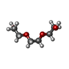
 Mass: 106.120 Da / Num. of mol.: 4 / Source method: obtained synthetically / Formula: C4H10O3
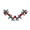
 Mass: 194.226 Da / Num. of mol.: 1 / Source method: obtained synthetically / Formula: C8H18O5 / Comment: precipitant*YM
 Mass: 18.015 Da / Num. of mol.: 916 / Source method: isolated from a natural source / Formula: H2O
 Sample preparation
 / Beamline: BL11-1 / Wavelength: 0.91837, 0.97901, 0.97926
 Processing