 Movie
Movie Controller


+ Open data

- Basic information
| Entry | Database: PDB / ID: 2pom | ||||||
|---|---|---|---|---|---|---|---|
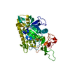
| Title | TAB1 with manganese ion | ||||||
 Components Components | Mitogen-activated protein kinase kinase kinase 7-interacting protein 1 | ||||||
 Keywords Keywords |  SIGNALING PROTEIN/METAL BINDING PROTEIN / pp2c-like domain / SIGNALING PROTEIN-METAL BINDING PROTEIN COMPLEX SIGNALING PROTEIN/METAL BINDING PROTEIN / pp2c-like domain / SIGNALING PROTEIN-METAL BINDING PROTEIN COMPLEX | ||||||
| Function / homology |  Function and homology information Function and homology informationcardiac septum development / coronary vasculature development / aorta development / protein serine/threonine phosphatase activity / mitogen-activated protein kinase p38 binding / non-canonical NF-kappaB signal transduction / enzyme activator activity / heart morphogenesis / IRAK2 mediated activation of TAK1 complex / Alpha-protein kinase 1 signaling pathway ...cardiac septum development / coronary vasculature development / aorta development / protein serine/threonine phosphatase activity / mitogen-activated protein kinase p38 binding / non-canonical NF-kappaB signal transduction / enzyme activator activity / heart morphogenesis / IRAK2 mediated activation of TAK1 complex / Alpha-protein kinase 1 signaling pathway / IRAK2 mediated activation of TAK1 complex upon TLR7/8 or 9 stimulation / TICAM1,TRAF6-dependent induction of TAK1 complex / TRAF6-mediated induction of TAK1 complex within TLR4 complex / protein serine/threonine kinase activator activity / transforming growth factor beta receptor signaling pathway / JNK (c-Jun kinases) phosphorylation and activation mediated by activated human TAK1 / TNFR1-induced NF-kappa-B signaling pathway / activated TAK1 mediates p38 MAPK activation / lung development / TAK1-dependent IKK and NF-kappa-B activation / NOD1/2 Signaling Pathway / positive regulation of protein serine/threonine kinase activity / CLEC7A (Dectin-1) signaling / FCERI mediated NF-kB activation / Interleukin-1 signaling / in utero embryonic development / positive regulation of MAPK cascade / molecular adaptor activity / endosome membrane / Ub-specific processing proteases / nuclear speck / protein-containing complex binding / SARS-CoV-2 activates/modulates innate and adaptive immune responses / endoplasmic reticulum / protein-containing complex / cytosol / cytoplasmSimilarity search - Function | ||||||
| Biological species |  Homo sapiens (human) Homo sapiens (human) | ||||||
| Method | X-RAY DIFFRACTION / SYNCHROTRON / MOLECULAR REPLACEMENT / Resolution: 2.27 Å | ||||||
 Authors Authors | Lin, S.C. | ||||||
 Citation Citation | Journal: Mol.Cell / Year: 2007 Title: XIAP Induces NF-kappaB Activation via the BIR1/TAB1 Interaction and BIR1 Dimerization. Authors: Lu, M. / Lin, S.C. / Huang, Y. / Kang, Y.J. / Rich, R. / Lo, Y.C. / Myszka, D. / Han, J. / Wu, H. | ||||||
| History |
|
- Structure visualization
| Structure viewer | Molecule: MolmilJmol/JSmol |
|---|
- Downloads & links
-Download
| PDBx/mmCIF format | 2pom.cif.gz | 82.2 KB | Display | PDBx/mmCIF format |
|---|---|---|---|---|
| PDB format | pdb2pom.ent.gz | 60.9 KB | Display | PDB format |
| PDBx/mmJSON format | 2pom.json.gz | Tree view | PDBx/mmJSON format | |
| Others |  Other downloads Other downloads |
-Validation report
| Arichive directory | https://data.pdbj.org/pub/pdb/validation_reports/po/2pomftp://data.pdbj.org/pub/pdb/validation_reports/po/2pom | HTTPS FTP |
|---|
-Related structure data


-Links
 PDBj
PDBj


- Assembly
| Deposited unit | 
| ||||||||
|---|---|---|---|---|---|---|---|---|---|
| 1 |
| ||||||||
| Unit cell |
| ||||||||
| Details | The biological assembly is a monomer. |
-Components
| #1: Protein | Mass: 40786.797 Da / Num. of mol.: 1 / Fragment: N-TERMINAL PP2C-LIKE DOMAIN, RESIDUES 1-370 Source method: isolated from a genetically manipulated source Source: (gene. exp.) Homo sapiens (human) / Gene: MAP3K7IP1, TAB1 / Plasmid: pGEX-4T3 / Species (production host): Escherichia coli / Production host:  Escherichia coli K12 (bacteria) / Strain (production host): K-12 / References: UniProt: Q15750 Escherichia coli K12 (bacteria) / Strain (production host): K-12 / References: UniProt: Q15750 |
|---|---|
| #2: Chemical | ChemComp-MN /   Mass: 54.938 Da / Num. of mol.: 1 / Source method: obtained synthetically / Formula: Mn Mass: 54.938 Da / Num. of mol.: 1 / Source method: obtained synthetically / Formula: Mn |
| #3: Water | ChemComp-HOH / Water Mass: 18.015 Da / Num. of mol.: 15 / Source method: isolated from a natural source / Formula: H2O Mass: 18.015 Da / Num. of mol.: 15 / Source method: isolated from a natural source / Formula: H2O |
-Experimental details
-Experiment
| Experiment | Method: X-RAY DIFFRACTION / Number of used crystals: 1 |
|---|
- Sample preparation
| Crystal | Density Matthews: 4.71 Å3/Da / Density % sol: 73.89 % |
|---|---|
| Crystal grow | Temperature: 295 K / Method: vapor diffusion, hanging drop / pH: 7.5 Details: 1.5M Li2SO4, 0.1M Hepes, 1mM MnCl2, pH 7.5, VAPOR DIFFUSION, HANGING DROP, temperature 295K |
-Data collection
| Diffraction | Mean temperature: 213 K | ||||||||||||||||||||||||||||||||||||||||||||||||||||||||||||||||||||||||||||||||||||||||||||||||||||||||||||||||||||||||||||||
|---|---|---|---|---|---|---|---|---|---|---|---|---|---|---|---|---|---|---|---|---|---|---|---|---|---|---|---|---|---|---|---|---|---|---|---|---|---|---|---|---|---|---|---|---|---|---|---|---|---|---|---|---|---|---|---|---|---|---|---|---|---|---|---|---|---|---|---|---|---|---|---|---|---|---|---|---|---|---|---|---|---|---|---|---|---|---|---|---|---|---|---|---|---|---|---|---|---|---|---|---|---|---|---|---|---|---|---|---|---|---|---|---|---|---|---|---|---|---|---|---|---|---|---|---|---|---|---|
| Diffraction source | Source: SYNCHROTRON / Site: NSLS  / Beamline: X4A / Wavelength: 0.97919 Å / Beamline: X4A / Wavelength: 0.97919 Å | ||||||||||||||||||||||||||||||||||||||||||||||||||||||||||||||||||||||||||||||||||||||||||||||||||||||||||||||||||||||||||||||
| Detector | Type: ADSC QUANTUM 4 / Detector: CCD / Date: Mar 6, 2006 | ||||||||||||||||||||||||||||||||||||||||||||||||||||||||||||||||||||||||||||||||||||||||||||||||||||||||||||||||||||||||||||||
| Radiation | Protocol: SINGLE WAVELENGTH / Monochromatic (M) / Laue (L): M / Scattering type: x-ray | ||||||||||||||||||||||||||||||||||||||||||||||||||||||||||||||||||||||||||||||||||||||||||||||||||||||||||||||||||||||||||||||
| Radiation wavelength | Wavelength: 0.97919 Å / Relative weight: 1 | ||||||||||||||||||||||||||||||||||||||||||||||||||||||||||||||||||||||||||||||||||||||||||||||||||||||||||||||||||||||||||||||
| Reflection | Resolution: 2.22→50 Å / Num. obs: 35865 / % possible obs: 95 % / Redundancy: 6.2 % / Rmerge(I) obs: 0.069 / Χ2: 1.159 / Net I/σ(I): 13.2 | ||||||||||||||||||||||||||||||||||||||||||||||||||||||||||||||||||||||||||||||||||||||||||||||||||||||||||||||||||||||||||||||
| Reflection shell |
|
- Processing
| Software |
| ||||||||||||||||||||||||||||||||||||||||||
|---|---|---|---|---|---|---|---|---|---|---|---|---|---|---|---|---|---|---|---|---|---|---|---|---|---|---|---|---|---|---|---|---|---|---|---|---|---|---|---|---|---|---|---|
| Refinement | Method to determine structure: MOLECULAR REPLACEMENT Starting model: The TAB1 model solved by ourselves, but Not deposited yet Resolution: 2.27→50 Å / Cross valid method: THROUGHOUT / σ(F): 0 / Stereochemistry target values: Engh & Huber
| ||||||||||||||||||||||||||||||||||||||||||
| Solvent computation | Bsol: 44.339 Å2 | ||||||||||||||||||||||||||||||||||||||||||
| Displacement parameters | Biso mean: 56.886 Å2
| ||||||||||||||||||||||||||||||||||||||||||
| Refinement step | Cycle: LAST / Resolution: 2.27→50 Å
| ||||||||||||||||||||||||||||||||||||||||||
| LS refinement shell |
| ||||||||||||||||||||||||||||||||||||||||||
| Xplor file |
|
