 Movie
Movie Controller

[English] 日本語
 Yorodumi
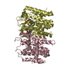
- PDB-2iqt: Crystal Structure of Fructose-Bisphosphate Aldolase, Class I from... -
+ Open data

- Basic information
| Entry | Database: PDB / ID: 2iqt | ||||||
|---|---|---|---|---|---|---|---|
| Title | Crystal Structure of Fructose-Bisphosphate Aldolase, Class I from Porphyromonas gingivalis | ||||||
 Components Components | Fructose-bisphosphate aldolase class 1 | ||||||
 Keywords Keywords |  LYASE / TIM berrel / Structural Genomics / PSI-2 / Protein Structure Initiative / Midwest Center for Structural Genomics / MCSG LYASE / TIM berrel / Structural Genomics / PSI-2 / Protein Structure Initiative / Midwest Center for Structural Genomics / MCSG | ||||||
| Function / homology |  Function and homology informationfructose-bisphosphate aldolase / fructose-bisphosphate aldolase activity / glycolytic process Function and homology informationfructose-bisphosphate aldolase / fructose-bisphosphate aldolase activity / glycolytic processSimilarity search - Function | ||||||
| Biological species |  Porphyromonas gingivalis (bacteria) Porphyromonas gingivalis (bacteria) | ||||||
| Method | X-RAY DIFFRACTION / SYNCHROTRON / SAD / Resolution: 2.46 Å | ||||||
 Authors Authors | Kim, Y. / Zhou, M. / Moy, S. / Joachimiak, A. / Midwest Center for Structural Genomics (MCSG) | ||||||
 Citation Citation | Journal: To be Published Title: Crystal Structure of Fructose-Bisphosphate Aldolase, Class I from Porphyromonas gingivalis Authors: Kim, Y. / Zhou, M. / Moy, S. / Joachimiak, A. | ||||||
| History |
|
- Structure visualization
| Structure viewer | Molecule: MolmilJmol/JSmol |
|---|
- Downloads & links
-Download
| PDBx/mmCIF format | 2iqt.cif.gz | 72.6 KB | Display | PDBx/mmCIF format |
|---|---|---|---|---|
| PDB format | pdb2iqt.ent.gz | 59.4 KB | Display | PDB format |
| PDBx/mmJSON format | 2iqt.json.gz | Tree view | PDBx/mmJSON format | |
| Others |  Other downloads Other downloads |
-Validation report
| Arichive directory | https://data.pdbj.org/pub/pdb/validation_reports/iq/2iqtftp://data.pdbj.org/pub/pdb/validation_reports/iq/2iqt | HTTPS FTP |
|---|
-Related structure data
| Similar structure data | |
|---|---|
| Other databases |


-Links
 PDBj
PDBj
- Assembly
| Deposited unit | 
| |||||||||
|---|---|---|---|---|---|---|---|---|---|---|
| 1 | 
| |||||||||
| Unit cell |
| |||||||||
| Components on special symmetry positions |
|
-Components
| #1: Protein | Mass: 33956.910 Da / Num. of mol.: 1 Source method: isolated from a genetically manipulated source Source: (gene. exp.) Porphyromonas gingivalis (bacteria) / Strain: W83 / Gene: fbaB / Plasmid: pMCSG7 / Species (production host): Escherichia coli / Production host: Escherichia coli BL21(DE3) (bacteria) / Strain (production host): BL21DE3 / References: UniProt: P60053, fructose-bisphosphate aldolase |
|---|---|
| #2: Water | ChemComp-HOH / Water Mass: 18.015 Da / Num. of mol.: 128 / Source method: isolated from a natural source / Formula: H2O Mass: 18.015 Da / Num. of mol.: 128 / Source method: isolated from a natural source / Formula: H2O |
-Experimental details
-Experiment
| Experiment | Method: X-RAY DIFFRACTION / Number of used crystals: 1 |
|---|
- Sample preparation
| Crystal | Density Matthews: 3.09 Å3/Da / Density % sol: 60.24 % |
|---|---|
| Crystal grow | Temperature: 289 K / Method: vapor diffusion, sitting drop / pH: 7 Details: 1.2 M Ammonium citrate tribasic, 100 mM BTP, pH 7.0, VAPOR DIFFUSION, SITTING DROP, temperature 289K |
-Data collection
| Diffraction | Mean temperature: 100 K |
|---|---|
| Diffraction source | Source: SYNCHROTRON / Site: APS  / Beamline: 19-BM / Wavelength: 0.97869 / Beamline: 19-BM / Wavelength: 0.97869 |
| Detector | Type: SBC-3 / Detector: CCD / Date: Dec 17, 2005 / Details: mirrors |
| Radiation | Monochromator: double crystal monochromator / Protocol: SINGLE WAVELENGTH / Monochromatic (M) / Laue (L): M / Scattering type: x-ray |
| Radiation wavelength | Wavelength: 0.97869 Å / Relative weight: 1 |
| Reflection | Resolution: 2.44→38.89 Å / Num. all: 16237 / Num. obs: 16237 / % possible obs: 99.7 % / Observed criterion σ(F): 0 / Observed criterion σ(I): 0 / Redundancy: 8.5 % / Rmerge(I) obs: 0.144 / Net I/σ(I): 7 |
| Reflection shell | Resolution: 2.44→2.53 Å / Redundancy: 6.1 % / Rmerge(I) obs: 0.427 / Mean I/σ(I) obs: 3.8 / Num. unique all: 1602 / % possible all: 99.8 |
- Processing
| Software |
| ||||||||||||||||||||||||||||||||||||||||||||||||||||||||||||||||||||||||||||||||||||||||||||||||||||||||||||||||||||||||||||||||||||||||||||||||||||||||||||||||||||||||||
|---|---|---|---|---|---|---|---|---|---|---|---|---|---|---|---|---|---|---|---|---|---|---|---|---|---|---|---|---|---|---|---|---|---|---|---|---|---|---|---|---|---|---|---|---|---|---|---|---|---|---|---|---|---|---|---|---|---|---|---|---|---|---|---|---|---|---|---|---|---|---|---|---|---|---|---|---|---|---|---|---|---|---|---|---|---|---|---|---|---|---|---|---|---|---|---|---|---|---|---|---|---|---|---|---|---|---|---|---|---|---|---|---|---|---|---|---|---|---|---|---|---|---|---|---|---|---|---|---|---|---|---|---|---|---|---|---|---|---|---|---|---|---|---|---|---|---|---|---|---|---|---|---|---|---|---|---|---|---|---|---|---|---|---|---|---|---|---|---|---|---|---|
| Refinement | Method to determine structure: SAD / Resolution: 2.46→38.89 Å / Cor.coef. Fo:Fc: 0.949 / Cor.coef. Fo:Fc free: 0.918 / SU B: 14.105 / SU ML: 0.166 / TLS residual ADP flag: LIKELY RESIDUAL / Cross valid method: THROUGHOUT / σ(F): 0 / σ(I): 0 / ESU R: 0.371 / ESU R Free: 0.251 / Stereochemistry target values: MAXIMUM LIKELIHOOD
| ||||||||||||||||||||||||||||||||||||||||||||||||||||||||||||||||||||||||||||||||||||||||||||||||||||||||||||||||||||||||||||||||||||||||||||||||||||||||||||||||||||||||||
| Solvent computation | Ion probe radii: 0.8 Å / Shrinkage radii: 0.8 Å / VDW probe radii: 1.4 Å / Solvent model: MASK | ||||||||||||||||||||||||||||||||||||||||||||||||||||||||||||||||||||||||||||||||||||||||||||||||||||||||||||||||||||||||||||||||||||||||||||||||||||||||||||||||||||||||||
| Displacement parameters | Biso mean: 35.452 Å2
| ||||||||||||||||||||||||||||||||||||||||||||||||||||||||||||||||||||||||||||||||||||||||||||||||||||||||||||||||||||||||||||||||||||||||||||||||||||||||||||||||||||||||||
| Refinement step | Cycle: LAST / Resolution: 2.46→38.89 Å
| ||||||||||||||||||||||||||||||||||||||||||||||||||||||||||||||||||||||||||||||||||||||||||||||||||||||||||||||||||||||||||||||||||||||||||||||||||||||||||||||||||||||||||
| Refine LS restraints |
| ||||||||||||||||||||||||||||||||||||||||||||||||||||||||||||||||||||||||||||||||||||||||||||||||||||||||||||||||||||||||||||||||||||||||||||||||||||||||||||||||||||||||||
| LS refinement shell | Resolution: 2.457→2.521 Å / Total num. of bins used: 20
| ||||||||||||||||||||||||||||||||||||||||||||||||||||||||||||||||||||||||||||||||||||||||||||||||||||||||||||||||||||||||||||||||||||||||||||||||||||||||||||||||||||||||||
| Refinement TLS params. | Method: refined / Origin x: 26.4417 Å / Origin y: 31.1258 Å / Origin z: 61.6001 Å
|
