 Movie
Movie Controller

[English] 日本語
 Yorodumi
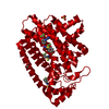
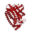
- PDB-2i19: T. Brucei farnesyl diphosphate synthase complexed with bisphosphonate -
+ Open data

- Basic information
| Entry | Database: PDB / ID: 2i19 | ||||||
|---|---|---|---|---|---|---|---|
| Title | T. Brucei farnesyl diphosphate synthase complexed with bisphosphonate | ||||||
 Components Components | Farnesyl pyrophosphate synthase Dimethylallyltranstransferase Dimethylallyltranstransferase | ||||||
 Keywords Keywords | TRANSFERASE / PROTEIN-BISPHOSPHONATE COMPLEX | ||||||
| Function / homology |  Function and homology information Function and homology informationtransferase activity, transferring alkyl or aryl (other than methyl) groups / isoprenoid biosynthetic process / metal ion bindingSimilarity search - Function | ||||||
| Biological species |  Trypanosoma brucei (eukaryote) Trypanosoma brucei (eukaryote) | ||||||
| Method | X-RAY DIFFRACTION / SYNCHROTRON / MOLECULAR REPLACEMENT / Resolution: 2.28 Å | ||||||
 Authors Authors | Cao, R. / Mao, J. / Gao, Y. / Robinson, H. / Odeh, S. / Goddard, A. / Oldfield, E. | ||||||
 Citation Citation | Journal: J.Am.Chem.Soc. / Year: 2006 Title: Solid-state NMR, crystallographic, and computational investigation of bisphosphonates and farnesyl diphosphate synthase-bisphosphonate complexes. Authors: Mao, J. / Mukherjee, S. / Zhang, Y. / Cao, R. / Sanders, J.M. / Song, Y. / Zhang, Y. / Meints, G.A. / Gao, Y.G. / Mukkamala, D. / Hudock, M.P. / Oldfield, E. | ||||||
| History |
|
- Structure visualization
| Structure viewer | Molecule: MolmilJmol/JSmol |
|---|
- Downloads & links
-Download
| PDBx/mmCIF format | 2i19.cif.gz | 166.7 KB | Display | PDBx/mmCIF format |
|---|---|---|---|---|
| PDB format | pdb2i19.ent.gz | 129.9 KB | Display | PDB format |
| PDBx/mmJSON format | 2i19.json.gz | Tree view | PDBx/mmJSON format | |
| Others |  Other downloads Other downloads |
-Validation report
| Arichive directory | https://data.pdbj.org/pub/pdb/validation_reports/i1/2i19ftp://data.pdbj.org/pub/pdb/validation_reports/i1/2i19 | HTTPS FTP |
|---|
-Related structure data
| Related structure data |  2ewgC 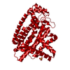 1yhkS S: Starting model for refinement C: citing same article ( |
|---|---|
| Similar structure data |


-Links
 PDBj
PDBj
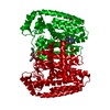
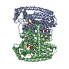
- Assembly
| Deposited unit | 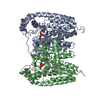
| ||||||||
|---|---|---|---|---|---|---|---|---|---|
| 1 |
| ||||||||
| Unit cell |
|
-Components
| #1: Protein | Dimethylallyltranstransferase Mass: 44475.738 Da / Num. of mol.: 2 Source method: isolated from a genetically manipulated source Source: (gene. exp.) Trypanosoma brucei (eukaryote) / Plasmid: pET-28a+ / Production host:  Escherichia coli (E. coli) / Strain (production host): BL21(DE3)-pLysS Escherichia coli (E. coli) / Strain (production host): BL21(DE3)-pLysSReferences: UniProt: Q86C09, (2E,6E)-farnesyl diphosphate synthase #2: Chemical | ChemComp-MG /   Mass: 24.305 Da / Num. of mol.: 6 / Source method: obtained synthetically / Formula: Mg Mass: 24.305 Da / Num. of mol.: 6 / Source method: obtained synthetically / Formula: Mg#3: Chemical |   Mass: 282.128 Da / Num. of mol.: 2 / Source method: obtained synthetically / Formula: C7H12N2O6P2 Mass: 282.128 Da / Num. of mol.: 2 / Source method: obtained synthetically / Formula: C7H12N2O6P2#4: Water | ChemComp-HOH / | Water Mass: 18.015 Da / Num. of mol.: 358 / Source method: isolated from a natural source / Formula: H2O Mass: 18.015 Da / Num. of mol.: 358 / Source method: isolated from a natural source / Formula: H2O |
|---|
-Experimental details
-Experiment
| Experiment | Method: X-RAY DIFFRACTION / Number of used crystals: 1 |
|---|
- Sample preparation
| Crystal | Density Matthews: 2.74 Å3/Da / Density % sol: 55.17 % |
|---|---|
| Crystal grow | Temperature: 298.15 K / Method: vapor diffusion, hanging drop / pH: 5.75 Details: 10% MPD, 0.1 AMMONIUM ACETATE, pH 5.75, VAPOR DIFFUSION, HANGING DROP, temperature 298.15K |
-Data collection
| Diffraction | Mean temperature: 123.2 K |
|---|---|
| Diffraction source | Source: SYNCHROTRON / Site: NSLS  / Beamline: X12C / Wavelength: 1.1 Å / Beamline: X12C / Wavelength: 1.1 Å |
| Detector | Type: ADSC QUANTUM 315 / Detector: CCD / Date: Apr 11, 2006 |
| Radiation | Monochromator: GRAPHITE / Protocol: SINGLE WAVELENGTH / Monochromatic (M) / Laue (L): M / Scattering type: x-ray |
| Radiation wavelength | Wavelength: 1.1 Å / Relative weight: 1 |
| Reflection | Resolution: 2.2→30 Å / Num. all: 41878 / Num. obs: 39495 / % possible obs: 91 % / Observed criterion σ(F): 0 / Redundancy: 6.6 % / Rmerge(I) obs: 0.073 / Net I/σ(I): 0.101 |
| Reflection shell | Resolution: 2.2→2.28 Å / Redundancy: 3.5 % / Rmerge(I) obs: 0.424 / % possible all: 56.5 |
- Processing
| Software |
| |||||||||||||||||||||||||||||||||
|---|---|---|---|---|---|---|---|---|---|---|---|---|---|---|---|---|---|---|---|---|---|---|---|---|---|---|---|---|---|---|---|---|---|---|
| Refinement | Method to determine structure: MOLECULAR REPLACEMENT Starting model: PDB ENTRY 1YHK Resolution: 2.28→30 Å / Num. parameters: 19711 / Num. restraintsaints: 24302 / Cross valid method: FREE R / σ(F): 0 / Stereochemistry target values: ENGH AND HUBER Details: ANISOTROPIC SCALING APPLIED BY THE METHOD OF PARKIN, MOEZZI & HOPE, J.APPL.CRYST.28(1995)53-56
| |||||||||||||||||||||||||||||||||
| Refine analyze | Num. disordered residues: 0 / Occupancy sum hydrogen: 0 / Occupancy sum non hydrogen: 6234 | |||||||||||||||||||||||||||||||||
| Refinement step | Cycle: LAST / Resolution: 2.28→30 Å
| |||||||||||||||||||||||||||||||||
| Refine LS restraints |
|
