 Movie
Movie Controller

[English] 日本語
 Yorodumi
- PDB-2hrk: Structural basis of yeast aminoacyl-tRNA synthetase complex forma... -
+ Open data

- Basic information
| Entry | Database: PDB / ID: 2hrk | ||||||
|---|---|---|---|---|---|---|---|
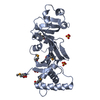
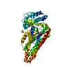
| Title | Structural basis of yeast aminoacyl-tRNA synthetase complex formation revealed by crystal structures of two binary sub-complexes | ||||||
 Components Components |
| ||||||
 Keywords Keywords | ligase/RNA Binding Protein /  protein complex protein interaction GST-fold / ligase-RNA Binding Protein COMPLEX protein complex protein interaction GST-fold / ligase-RNA Binding Protein COMPLEX | ||||||
| Function / homology |  Function and homology information Function and homology informationmethionyl glutamyl tRNA synthetase complex / glutamate-tRNA ligase / glutamate-tRNA ligase activity / glutamyl-tRNA aminoacylation / methionyl-tRNA aminoacylation / tRNA aminoacylation for protein translation / phosphatidylinositol-3-phosphate binding / phosphatidylinositol-3,5-bisphosphate binding / enzyme activator activity / cytoplasmic stress granule ...methionyl glutamyl tRNA synthetase complex / glutamate-tRNA ligase / glutamate-tRNA ligase activity / glutamyl-tRNA aminoacylation / methionyl-tRNA aminoacylation / tRNA aminoacylation for protein translation / phosphatidylinositol-3-phosphate binding / phosphatidylinositol-3,5-bisphosphate binding / enzyme activator activity / cytoplasmic stress granule / tRNA binding / mRNA binding / mitochondrion / ATP binding / cytosol / cytoplasmSimilarity search - Function | ||||||
| Biological species |  Saccharomyces cerevisiae (brewer's yeast) Saccharomyces cerevisiae (brewer's yeast) | ||||||
| Method | X-RAY DIFFRACTION / MOLECULAR REPLACEMENT / Resolution: 2.05 Å | ||||||
 Authors Authors | Simader, H. / Suck, D. | ||||||
 Citation Citation | Journal: Nucleic Acids Res. / Year: 2006 Title: Structural basis of yeast aminoacyl-tRNA synthetase complex formation revealed by crystal structures of two binary sub-complexes. Authors: Simader, H. / Hothorn, M. / Kohler, C. / Basquin, J. / Simos, G. / Suck, D. | ||||||
| History |
|
- Structure visualization
| Structure viewer | Molecule: MolmilJmol/JSmol |
|---|
- Downloads & links
-Download
| PDBx/mmCIF format | 2hrk.cif.gz | 77.9 KB | Display | PDBx/mmCIF format |
|---|---|---|---|---|
| PDB format | pdb2hrk.ent.gz | 56.5 KB | Display | PDB format |
| PDBx/mmJSON format | 2hrk.json.gz | Tree view | PDBx/mmJSON format | |
| Others |  Other downloads Other downloads |
-Validation report
| Arichive directory | https://data.pdbj.org/pub/pdb/validation_reports/hr/2hrkftp://data.pdbj.org/pub/pdb/validation_reports/hr/2hrk | HTTPS FTP |
|---|
-Related structure data
| Related structure data | 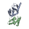 2hsmC  2hsnC  2hqtS  2hraS S: Starting model for refinement C: citing same article ( |
|---|---|
| Similar structure data |


-Links
 PDBj
PDBj


- Assembly
| Deposited unit | 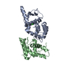
| ||||||||
|---|---|---|---|---|---|---|---|---|---|
| 1 |
| ||||||||
| Unit cell |
| ||||||||
| Details | The biological unit is the binary complex. The asymmetric unit contains one biological unit. |
-Components
| #1: Protein | Mass: 22765.432 Da / Num. of mol.: 1 / Fragment: Residues 1-207 Source method: isolated from a genetically manipulated source Source: (gene. exp.) Saccharomyces cerevisiae (brewer's yeast)Gene: GUS1 / Plasmid: pETm-derivative / Production host:  Escherichia coli (E. coli) / Strain (production host): BL21 DE3 Star / References: UniProt: P46655, glutamate-tRNA ligase Escherichia coli (E. coli) / Strain (production host): BL21 DE3 Star / References: UniProt: P46655, glutamate-tRNA ligase |
|---|---|
| #2: Protein | Mass: 14002.878 Da / Num. of mol.: 1 / Fragment: Residues 1-122 Source method: isolated from a genetically manipulated source Source: (gene. exp.) Saccharomyces cerevisiae (brewer's yeast)Gene: ARC1 / Plasmid: pETm-derivative / Production host: Escherichia coli (E. coli) / Strain (production host): BL21 DE3 Star / References: UniProt: P46672 |
| #3: Chemical | ChemComp-CL / Chloride  Mass: 35.453 Da / Num. of mol.: 1 / Source method: obtained synthetically / Formula: Cl Mass: 35.453 Da / Num. of mol.: 1 / Source method: obtained synthetically / Formula: Cl |
| #4: Water | ChemComp-HOH / Water Mass: 18.015 Da / Num. of mol.: 274 / Source method: isolated from a natural source / Formula: H2O Mass: 18.015 Da / Num. of mol.: 274 / Source method: isolated from a natural source / Formula: H2O |
-Experimental details
-Experiment
| Experiment | Method: X-RAY DIFFRACTION / Number of used crystals: 1 |
|---|
- Sample preparation
| Crystal | Density Matthews: 2.2 Å3/Da / Density % sol: 44.08 % |
|---|---|
| Crystal grow | Temperature: 293 K / Method: vapor diffusion, hanging drop / pH: 7.2 Details: 30-35 % PEG 3350, 0.3-0.5 M NaSCN, pH 7.2, VAPOR DIFFUSION, HANGING DROP, temperature 293K |
-Data collection
| Diffraction | Mean temperature: 100 K |
|---|---|
| Diffraction source | Source: ROTATING ANODE / Type: RIGAKU / Wavelength: 1.54179 Å |
| Detector | Type: MAR scanner 345 mm plate / Detector: IMAGE PLATE / Date: Jan 29, 2006 / Details: Osmic VariMax Multilayer |
| Radiation | Monochromator: Osmic mirrors / Protocol: SINGLE WAVELENGTH / Monochromatic (M) / Laue (L): M / Scattering type: x-ray |
| Radiation wavelength | Wavelength: 1.54179 Å / Relative weight: 1 |
| Reflection | Resolution: 2.05→45 Å / Num. obs: 20000 / % possible obs: 99.9 % / Observed criterion σ(F): -3 / Observed criterion σ(I): -3 / Redundancy: 4.1 % / Rsym value: 0.038 / Net I/σ(I): 27.45 |
| Reflection shell | Resolution: 2.05→2.1 Å / Redundancy: 4 % / Mean I/σ(I) obs: 5.75 / Num. unique all: 1396 / Rsym value: 0.28 / % possible all: 100 |
- Processing
| Software |
| ||||||||||||||||||||||||||||||||||||||||||||||||||||||||||||||||||||||||||||||||||||||||||||||||||||||||||||||||||||||||||||||||||
|---|---|---|---|---|---|---|---|---|---|---|---|---|---|---|---|---|---|---|---|---|---|---|---|---|---|---|---|---|---|---|---|---|---|---|---|---|---|---|---|---|---|---|---|---|---|---|---|---|---|---|---|---|---|---|---|---|---|---|---|---|---|---|---|---|---|---|---|---|---|---|---|---|---|---|---|---|---|---|---|---|---|---|---|---|---|---|---|---|---|---|---|---|---|---|---|---|---|---|---|---|---|---|---|---|---|---|---|---|---|---|---|---|---|---|---|---|---|---|---|---|---|---|---|---|---|---|---|---|---|---|---|
| Refinement | Method to determine structure: MOLECULAR REPLACEMENT Starting model: PDB entry 2HRA PDB entry 2HQT Resolution: 2.05→45 Å / Cor.coef. Fo:Fc: 0.947 / Cor.coef. Fo:Fc free: 0.904 / SU B: 5.252 / SU ML: 0.144 / Cross valid method: THROUGHOUT / σ(F): -3 / ESU R: 0.221 / ESU R Free: 0.198 / Stereochemistry target values: MAXIMUM LIKELIHOOD / Details: HYDROGENS HAVE BEEN ADDED IN THE RIDING POSITIONS
| ||||||||||||||||||||||||||||||||||||||||||||||||||||||||||||||||||||||||||||||||||||||||||||||||||||||||||||||||||||||||||||||||||
| Solvent computation | Ion probe radii: 0.8 Å / Shrinkage radii: 0.8 Å / VDW probe radii: 1.2 Å / Solvent model: BABINET MODEL WITH MASK | ||||||||||||||||||||||||||||||||||||||||||||||||||||||||||||||||||||||||||||||||||||||||||||||||||||||||||||||||||||||||||||||||||
| Displacement parameters | Biso mean: 26.461 Å2
| ||||||||||||||||||||||||||||||||||||||||||||||||||||||||||||||||||||||||||||||||||||||||||||||||||||||||||||||||||||||||||||||||||
| Refinement step | Cycle: LAST / Resolution: 2.05→45 Å
| ||||||||||||||||||||||||||||||||||||||||||||||||||||||||||||||||||||||||||||||||||||||||||||||||||||||||||||||||||||||||||||||||||
| Refine LS restraints |
| ||||||||||||||||||||||||||||||||||||||||||||||||||||||||||||||||||||||||||||||||||||||||||||||||||||||||||||||||||||||||||||||||||
| LS refinement shell | Resolution: 2.05→2.103 Å / Total num. of bins used: 20
|
