 Movie
Movie Controller

[English] 日本語
 Yorodumi
- PDB-2f8z: Crystal structure of human FPPS in complex with zoledronate and i... -
+ Open data

- Basic information
| Entry | Database: PDB / ID: 2f8z | ||||||
|---|---|---|---|---|---|---|---|
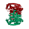
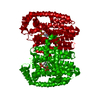
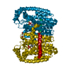
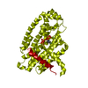
| Title | Crystal structure of human FPPS in complex with zoledronate and isopentenyl diphosphate | ||||||
 Components Components | Farnesyl Diphosphate Synthase | ||||||
 Keywords Keywords |  TRANSFERASE / Mevalonate pathway / isoprene biosynthesis / cholesterol biosynthesis / bisphosphonate inhibitor TRANSFERASE / Mevalonate pathway / isoprene biosynthesis / cholesterol biosynthesis / bisphosphonate inhibitor | ||||||
| Function / homology |  Function and homology information Function and homology informationgeranyl diphosphate biosynthetic process / dimethylallyltranstransferase / farnesyl diphosphate biosynthetic process / (2E,6E)-farnesyl diphosphate synthase / geranyltranstransferase activity / dimethylallyltranstransferase activity / Cholesterol biosynthesis / cholesterol biosynthetic process / Activation of gene expression by SREBF (SREBP) / RNA binding ...geranyl diphosphate biosynthetic process / dimethylallyltranstransferase / farnesyl diphosphate biosynthetic process / (2E,6E)-farnesyl diphosphate synthase / geranyltranstransferase activity / dimethylallyltranstransferase activity / Cholesterol biosynthesis / cholesterol biosynthetic process / Activation of gene expression by SREBF (SREBP) / RNA binding / nucleoplasm / metal ion binding / cytosol / cytoplasmSimilarity search - Function | ||||||
| Biological species |  Homo sapiens (human) Homo sapiens (human) | ||||||
| Method | X-RAY DIFFRACTION / SYNCHROTRON / FOURIER SYNTHESIS / Resolution: 2.6 Å | ||||||
 Authors Authors | Rondeau, J.-M. / Bitsch, F. / Bourgier, E. / Geiser, M. / Hemmig, R. / Kroemer, M. / Lehmann, S. / Ramage, P. / Rieffel, S. / Strauss, A. ...Rondeau, J.-M. / Bitsch, F. / Bourgier, E. / Geiser, M. / Hemmig, R. / Kroemer, M. / Lehmann, S. / Ramage, P. / Rieffel, S. / Strauss, A. / Green, J.R. / Jahnke, W. | ||||||
 Citation Citation | Journal: Chemmedchem / Year: 2006 Title: Structural basis for the exceptional in vivo efficacy of bisphosphonate drugs. Authors: Rondeau, J.M. / Bitsch, F. / Bourgier, E. / Geiser, M. / Hemmig, R. / Kroemer, M. / Lehmann, S. / Ramage, P. / Rieffel, S. / Strauss, A. / Green, J.R. / Jahnke, W. | ||||||
| History |
|
- Structure visualization
| Structure viewer | Molecule: MolmilJmol/JSmol |
|---|
- Downloads & links
-Download
| PDBx/mmCIF format | 2f8z.cif.gz | 88.4 KB | Display | PDBx/mmCIF format |
|---|---|---|---|---|
| PDB format | pdb2f8z.ent.gz | 65.2 KB | Display | PDB format |
| PDBx/mmJSON format | 2f8z.json.gz | Tree view | PDBx/mmJSON format | |
| Others |  Other downloads Other downloads |
-Validation report
| Arichive directory | https://data.pdbj.org/pub/pdb/validation_reports/f8/2f8zftp://data.pdbj.org/pub/pdb/validation_reports/f8/2f8z | HTTPS FTP |
|---|
-Related structure data
| Related structure data | 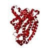 2f7mC 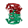 2f89C  2f8cSC 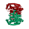 2f92C  2f94C  2f9kC C: citing same article ( S: Starting model for refinement |
|---|---|
| Similar structure data |


-Links
 PDBj
PDBj

- Assembly
| Deposited unit | 
| ||||||||
|---|---|---|---|---|---|---|---|---|---|
| 1 | 
| ||||||||
| Unit cell |
| ||||||||
| Details | THE BIOLOGICAL ASSEMBLY IS A HOMODIMER |
-Components
| #1: Protein | Mass: 40183.855 Da / Num. of mol.: 1 / Fragment: Residues 6-353 Source method: isolated from a genetically manipulated source Details: Includes: Dimethylallyltranstransferase; Geranyltranstransferase Source: (gene. exp.) Homo sapiens (human) / Gene: FDPS, FPS, KIAA1293 / Plasmid: pET28 / Production host:  Escherichia coli (E. coli) / Strain (production host): BL21 TUNER(DE3) Escherichia coli (E. coli) / Strain (production host): BL21 TUNER(DE3)References: UniProt: P14324, dimethylallyltranstransferase, (2E,6E)-farnesyl diphosphate synthase | ||||||
|---|---|---|---|---|---|---|---|
| #2: Chemical |   Mass: 24.305 Da / Num. of mol.: 3 / Source method: obtained synthetically / Formula: Mg Mass: 24.305 Da / Num. of mol.: 3 / Source method: obtained synthetically / Formula: Mg#3: Chemical | ChemComp-IPE / | Isopentenyl pyrophosphate  Mass: 246.092 Da / Num. of mol.: 1 / Source method: obtained synthetically / Formula: C5H12O7P2 Mass: 246.092 Da / Num. of mol.: 1 / Source method: obtained synthetically / Formula: C5H12O7P2#4: Chemical | ChemComp-ZOL / | Zoledronic acid  Mass: 272.090 Da / Num. of mol.: 1 / Source method: obtained synthetically / Formula: C5H10N2O7P2 / Comment: medication*YM Mass: 272.090 Da / Num. of mol.: 1 / Source method: obtained synthetically / Formula: C5H10N2O7P2 / Comment: medication*YM#5: Water | ChemComp-HOH / | Water Mass: 18.015 Da / Num. of mol.: 61 / Source method: isolated from a natural source / Formula: H2O Mass: 18.015 Da / Num. of mol.: 61 / Source method: isolated from a natural source / Formula: H2O |
-Experimental details
-Experiment
| Experiment | Method: X-RAY DIFFRACTION / Number of used crystals: 1 |
|---|
- Sample preparation
| Crystal | Density Matthews: 2.57 Å3/Da / Density % sol: 52.16 % |
|---|---|
| Crystal grow | Temperature: 292 K / Method: vapor diffusion, hanging drop / pH: 4.7 Details: 1.2M sodium potassium phosphate, 25% glycerol, pH 4.7, VAPOR DIFFUSION, HANGING DROP, temperature 292K |
-Data collection
| Diffraction | Mean temperature: 95 K |
|---|---|
| Diffraction source | Source: SYNCHROTRON / Site: SLS  / Beamline: X06SA / Wavelength: 1.00033 Å / Beamline: X06SA / Wavelength: 1.00033 Å |
| Detector | Type: MARRESEARCH / Detector: CCD / Date: Jan 29, 2004 |
| Radiation | Monochromator: SAGITALLY FOCUSED Si(111) / Protocol: SINGLE WAVELENGTH / Monochromatic (M) / Laue (L): M / Scattering type: x-ray |
| Radiation wavelength | Wavelength: 1.00033 Å / Relative weight: 1 |
| Reflection | Resolution: 2.6→100 Å / Num. all: 13248 / Num. obs: 13248 / % possible obs: 99.9 % / Observed criterion σ(F): 0 / Observed criterion σ(I): 0 / Redundancy: 7.7 % / Biso Wilson estimate: 62.5 Å2 / Rmerge(I) obs: 0.072 / Χ2: 1.035 / Net I/σ(I): 8.8 |
| Reflection shell | Resolution: 2.6→2.69 Å / Rmerge(I) obs: 0.481 / Num. unique all: 1282 / Χ2: 0.649 / % possible all: 99.8 |
- Processing
| Software |
| ||||||||||||||||||||||||||||||||||||
|---|---|---|---|---|---|---|---|---|---|---|---|---|---|---|---|---|---|---|---|---|---|---|---|---|---|---|---|---|---|---|---|---|---|---|---|---|---|
| Refinement | Method to determine structure: FOURIER SYNTHESIS Starting model: PDB ENTRY 2F8C Resolution: 2.6→56.71 Å / Rfactor Rfree error: 0.008 / Data cutoff high absF: 18221610 / Data cutoff low absF: 0 / Isotropic thermal model: RESTRAINED / Cross valid method: THROUGHOUT / σ(F): 0 / Stereochemistry target values: Engh & Huber
| ||||||||||||||||||||||||||||||||||||
| Solvent computation | Solvent model: FLAT MODEL / Bsol: 55.171 Å2 / ksol: 0.368 e/Å3 | ||||||||||||||||||||||||||||||||||||
| Displacement parameters | Biso mean: 59.9 Å2
| ||||||||||||||||||||||||||||||||||||
| Refine analyze |
| ||||||||||||||||||||||||||||||||||||
| Refinement step | Cycle: LAST / Resolution: 2.6→56.71 Å
| ||||||||||||||||||||||||||||||||||||
| Refine LS restraints |
| ||||||||||||||||||||||||||||||||||||
| LS refinement shell | Resolution: 2.6→2.76 Å / Rfactor Rfree error: 0.036 / Total num. of bins used: 6
| ||||||||||||||||||||||||||||||||||||
| Xplor file |
|
