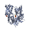
Entry Database : PDB / ID : 2.0E+88 Title Crystal structure of the human Hsp70 ATPase domain in the apo form Heat shock 70kDa protein 1A Keywords / / / / / Function / homology Function Domain/homology Component
/ / / / / / / / / / / / / / / / / / / / / / / / / / / / / / / / / / / / / / / / / / / / / / / / / / / / / / / / / / / / / / / / / / / / / / / / / / / / / / / / / / / / / / / / / / / / / / / / / / / / / / / / / / / / / / / / / / / / / / / / / / / / / Biological species Homo sapiens (human)Method / / / Resolution : 1.8 Å Authors Shida, M. / Ishii, R. / Takagi, T. / Kishishita, S. / Shirouzu, M. / Yokoyama, S. / RIKEN Structural Genomics/Proteomics Initiative (RSGI) Journal : Acta Crystallogr.,Sect.D / Year : 2010Title : Direct inter-subdomain interactions switch between the closed and open forms of the Hsp70 nucleotide-binding domain in the nucleotide-free state.Authors : Shida, M. / Arakawa, A. / Ishii, R. / Kishishita, S. / Takagi, T. / Kukimoto-Niino, M. / Sugano, S. / Tanaka, A. / Shirouzu, M. / Yokoyama, S. History Deposition Jan 19, 2007 Deposition site / Processing site Revision 1.0 Jan 22, 2008 Provider / Type Revision 1.1 Jul 13, 2011 Group Revision 1.2 Oct 25, 2023 Group Data collection / Database references ... Data collection / Database references / Derived calculations / Refinement description Category chem_comp_atom / chem_comp_bond ... chem_comp_atom / chem_comp_bond / database_2 / pdbx_initial_refinement_model / struct_ref_seq_dif / struct_site Item _database_2.pdbx_DOI / _database_2.pdbx_database_accession ... _database_2.pdbx_DOI / _database_2.pdbx_database_accession / _struct_ref_seq_dif.details / _struct_site.pdbx_auth_asym_id / _struct_site.pdbx_auth_comp_id / _struct_site.pdbx_auth_seq_id
Show all Show less
 Movie
Movie Controller

 Yorodumi
 Open data
 Basic information
 Components
 HSPA1A
  Keywords
 Function and homology information
 Authors
 Citation
 Structure visualization
 Downloads & links
 Other downloads


 PDBj
PDBj


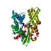
 Assembly


 Mass: 65.409 Da / Num. of mol.: 1 / Source method: obtained synthetically / Formula: Zn
 Mass: 18.015 Da / Num. of mol.: 416 / Source method: isolated from a natural source / Formula: H2O
 Sample preparation
 / Beamline: BL-5A / Wavelength: 1 Å
 Processing