 Movie
Movie Controller

[English] 日本語
 Yorodumi
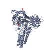
- PDB-2cbj: Structure of the Clostridium perfringens NagJ family 84 glycoside... -
+ Open data

- Basic information
| Entry | Database: PDB / ID: 2cbj | ||||||
|---|---|---|---|---|---|---|---|
| Title | Structure of the Clostridium perfringens NagJ family 84 glycoside hydrolase, a homologue of human O-GlcNAcase in complex with PUGNAc | ||||||
 Components Components | HYALURONIDASE | ||||||
 Keywords Keywords | HYDROLASE / O-GLCNAC / FAMILY 84 GLYCOSIDE HYDROLASES / GLYCOSIDE HYDROLASE / HYALURONIDASES / CARBOHYDRATES | ||||||
| Function / homology |  Function and homology information Function and homology informationorganonitrogen compound metabolic process / carbohydrate derivative metabolic process / protein O-GlcNAcase / : / : / [protein]-3-O-(N-acetyl-D-glucosaminyl)-L-serine/L-threonine O-N-acetyl-alpha-D-glucosaminase activity / protein deglycosylation / polysaccharide catabolic process / beta-N-acetylglucosaminidase activity / carbohydrate binding / carbohydrate metabolic processSimilarity search - Function | ||||||
| Biological species |   CLOSTRIDIUM PERFRINGENS (bacteria) CLOSTRIDIUM PERFRINGENS (bacteria) | ||||||
| Method | X-RAY DIFFRACTION / MOLECULAR REPLACEMENT / Resolution: 2.35 Å | ||||||
 Authors Authors | Rao, F.V. / Dorfmueller, H.C. / Villa, F. / Allwood, M. / Eggleston, I.M. / van Aalten, D.M.F. | ||||||
 Citation Citation | Journal: EMBO J. / Year: 2006 Title: Structural insights into the mechanism and inhibition of eukaryotic O-GlcNAc hydrolysis. Authors: Rao, F.V. / Dorfmueller, H.C. / Villa, F. / Allwood, M. / Eggleston, I.M. / van Aalten, D.M. | ||||||
| History |
| ||||||
| Remark 700 | SHEET DETERMINATION METHOD: DSSP THE SHEETS PRESENTED AS "AC" IN EACH CHAIN ON SHEET RECORDS BELOW ... SHEET DETERMINATION METHOD: DSSP THE SHEETS PRESENTED AS "AC" IN EACH CHAIN ON SHEET RECORDS BELOW IS ACTUALLY AN 8-STRANDED BARREL THIS IS REPRESENTED BY A 9-STRANDED SHEET IN WHICH THE FIRST AND LAST STRANDS ARE IDENTICAL. THE SHEETS PRESENTED AS "BC" IN EACH CHAIN ON SHEET RECORDS BELOW IS ACTUALLY AN 8-STRANDED BARREL THIS IS REPRESENTED BY A 9-STRANDED SHEET IN WHICH THE FIRST AND LAST STRANDS ARE IDENTICAL. |
- Structure visualization
| Structure viewer | Molecule: MolmilJmol/JSmol |
|---|
- Downloads & links
-Download
| PDBx/mmCIF format | 2cbj.cif.gz | 469.7 KB | Display | PDBx/mmCIF format |
|---|---|---|---|---|
| PDB format | pdb2cbj.ent.gz | 387.7 KB | Display | PDB format |
| PDBx/mmJSON format | 2cbj.json.gz | Tree view | PDBx/mmJSON format | |
| Others |  Other downloads Other downloads |
-Validation report
| Arichive directory | https://data.pdbj.org/pub/pdb/validation_reports/cb/2cbjftp://data.pdbj.org/pub/pdb/validation_reports/cb/2cbj | HTTPS FTP |
|---|
-Related structure data
| Related structure data |  2cbiSC S: Starting model for refinement C: citing same article ( |
|---|---|
| Similar structure data |


-Links
 PDBj
PDBj


- Assembly
| Deposited unit | 
| |||||||||
|---|---|---|---|---|---|---|---|---|---|---|
| 1 | 
| |||||||||
| 2 | 
| |||||||||
| Unit cell |
| |||||||||
| Components on special symmetry positions |
|
-Components
| #1: Protein | Mass: 66688.711 Da / Num. of mol.: 2 / Fragment: RESIDUES 31-624 Source method: isolated from a genetically manipulated source Source: (gene. exp.) CLOSTRIDIUM PERFRINGENS (bacteria)Description: DEOXYRIBONUCLEIC ACID FROM CLOSTRIDIUM PERFRINGENS (C. WELCHII) SIGMA (D5139) Plasmid: PGEX6P-1 / Production host: Escherichia coli BL21(DE3) (bacteria) / Variant (production host): pLysSReferences: UniProt: Q8XL08, UniProt: Q0TR53*PLUS, hyaluronoglucosaminidase#2: Chemical | PUGNAc  Mass: 353.327 Da / Num. of mol.: 2 / Source method: obtained synthetically / Formula: C15H19N3O7 / Comment: inhibitor*YM Mass: 353.327 Da / Num. of mol.: 2 / Source method: obtained synthetically / Formula: C15H19N3O7 / Comment: inhibitor*YM#3: Chemical | Chloride  Mass: 35.453 Da / Num. of mol.: 2 / Source method: obtained synthetically / Formula: Cl Mass: 35.453 Da / Num. of mol.: 2 / Source method: obtained synthetically / Formula: Cl#4: Water | ChemComp-HOH / | Water Mass: 18.015 Da / Num. of mol.: 280 / Source method: isolated from a natural source / Formula: H2O Mass: 18.015 Da / Num. of mol.: 280 / Source method: isolated from a natural source / Formula: H2OSequence details | ACCORDING TO THE AUTHORS THE CONFLICTS IN SEQADV ARE ALL VARIANTS | |
|---|
-Experimental details
-Experiment
| Experiment | Method: X-RAY DIFFRACTION / Number of used crystals: 1 |
|---|
- Sample preparation
| Crystal | Density Matthews: 2.69 Å3/Da / Density % sol: 53.86 % |
|---|---|
| Crystal grow | pH: 6.5 Details: 0.2 M AMMONIUM SULFATE, 0.1 M SODIUM CACODYLATE PH 6.5 AND 30 % PEG 8000) AND 0.25 MICROLITERS OF 40 % V/V GAMMA-BUTYROLACTONE ADDED TO A 1 PLUS 1 MICROLITER DROP |
-Data collection
| Diffraction | Mean temperature: 157 K |
|---|---|
| Diffraction source | Source: ROTATING ANODE / Type: RIGAKU MICROMAX-007 / Wavelength: 1.5418 |
| Detector | Type: RIGAKU RAXIS IV / Detector: IMAGE PLATE / Date: Nov 9, 2005 / Details: MIRRORS |
| Radiation | Protocol: SINGLE WAVELENGTH / Monochromatic (M) / Laue (L): M / Scattering type: x-ray |
| Radiation wavelength | Wavelength: 1.5418 Å / Relative weight: 1 |
| Reflection | Resolution: 2.35→20 Å / Num. obs: 56462 / % possible obs: 93.7 % / Observed criterion σ(I): 2 / Redundancy: 3.9 % / Rmerge(I) obs: 0.08 / Net I/σ(I): 15.8 |
| Reflection shell | Resolution: 2.35→2.43 Å / Redundancy: 3.6 % / Rmerge(I) obs: 0.61 / Mean I/σ(I) obs: 2.6 / % possible all: 96 |
- Processing
| Software |
| ||||||||||||||||||||||||||||||||||||||||||||||||||||||||||||||||||||||||||||||||||||||||||||||||||||||||||||||||||||||||||||||||||||||||||||||||||||||||||||||||||||||||||||||||||||||
|---|---|---|---|---|---|---|---|---|---|---|---|---|---|---|---|---|---|---|---|---|---|---|---|---|---|---|---|---|---|---|---|---|---|---|---|---|---|---|---|---|---|---|---|---|---|---|---|---|---|---|---|---|---|---|---|---|---|---|---|---|---|---|---|---|---|---|---|---|---|---|---|---|---|---|---|---|---|---|---|---|---|---|---|---|---|---|---|---|---|---|---|---|---|---|---|---|---|---|---|---|---|---|---|---|---|---|---|---|---|---|---|---|---|---|---|---|---|---|---|---|---|---|---|---|---|---|---|---|---|---|---|---|---|---|---|---|---|---|---|---|---|---|---|---|---|---|---|---|---|---|---|---|---|---|---|---|---|---|---|---|---|---|---|---|---|---|---|---|---|---|---|---|---|---|---|---|---|---|---|---|---|---|---|
| Refinement | Method to determine structure: MOLECULAR REPLACEMENT Starting model: PDB ENTRY 2CBI Resolution: 2.35→20 Å / Cor.coef. Fo:Fc: 0.953 / Cor.coef. Fo:Fc free: 0.925 / SU B: 15.357 / SU ML: 0.185 / Cross valid method: THROUGHOUT / ESU R: 0.345 / ESU R Free: 0.248 / Stereochemistry target values: MAXIMUM LIKELIHOOD Details: HYDROGENS HAVE BEEN ADDED IN THE RIDING POSITIONS. RESIDUES 31 TO 39 ARE DISORDERED AND HAVE NOT BEEN MODELLED
| ||||||||||||||||||||||||||||||||||||||||||||||||||||||||||||||||||||||||||||||||||||||||||||||||||||||||||||||||||||||||||||||||||||||||||||||||||||||||||||||||||||||||||||||||||||||
| Solvent computation | Ion probe radii: 0.8 Å / Shrinkage radii: 0.8 Å / VDW probe radii: 1.2 Å / Solvent model: BABINET MODEL WITH MASK | ||||||||||||||||||||||||||||||||||||||||||||||||||||||||||||||||||||||||||||||||||||||||||||||||||||||||||||||||||||||||||||||||||||||||||||||||||||||||||||||||||||||||||||||||||||||
| Displacement parameters | Biso mean: 42.28 Å2
| ||||||||||||||||||||||||||||||||||||||||||||||||||||||||||||||||||||||||||||||||||||||||||||||||||||||||||||||||||||||||||||||||||||||||||||||||||||||||||||||||||||||||||||||||||||||
| Refinement step | Cycle: LAST / Resolution: 2.35→20 Å
| ||||||||||||||||||||||||||||||||||||||||||||||||||||||||||||||||||||||||||||||||||||||||||||||||||||||||||||||||||||||||||||||||||||||||||||||||||||||||||||||||||||||||||||||||||||||
| Refine LS restraints |
|