 Movie
Movie Controller


+ Open data

- Basic information
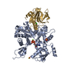
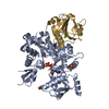
| Entry | Database: PDB / ID: 1yag | ||||||
|---|---|---|---|---|---|---|---|
| Title | STRUCTURE OF THE YEAST ACTIN-HUMAN GELSOLIN SEGMENT 1 COMPLEX | ||||||
 Components Components |
| ||||||
 Keywords Keywords |  CONTRACTILE PROTEIN / COMPLEX / ACTIN / GELSOLIN CONTRACTILE PROTEIN / COMPLEX / ACTIN / GELSOLIN | ||||||
| Function / homology |  Function and homology information Function and homology informationRHOA GTPase cycle / cellular bud neck contractile ring / mitotic actomyosin contractile ring contraction / RHO GTPases activate IQGAPs / RHO GTPases Activate WASPs and WAVEs / Regulation of actin dynamics for phagocytic cup formation / ascospore wall assembly / vacuole inheritance / striated muscle atrophy / regulation of establishment of T cell polarity ...RHOA GTPase cycle / cellular bud neck contractile ring / mitotic actomyosin contractile ring contraction / RHO GTPases activate IQGAPs / RHO GTPases Activate WASPs and WAVEs / Regulation of actin dynamics for phagocytic cup formation / ascospore wall assembly / vacuole inheritance / striated muscle atrophy / regulation of establishment of T cell polarity / regulation of plasma membrane raft polarization / regulation of receptor clustering / renal protein absorption / positive regulation of keratinocyte apoptotic process / positive regulation of protein processing in phagocytic vesicle / positive regulation of actin nucleation / phosphatidylinositol 3-kinase catalytic subunit binding / positive regulation of cysteine-type endopeptidase activity involved in apoptotic signaling pathway / actin cap / actin cortical patch / sequestering of actin monomers / regulation of podosome assembly / Swr1 complex / myosin II binding / Platelet degranulation / : / negative regulation of viral entry into host cell / actin filament severing / Ino80 complex / actin filament capping / barbed-end actin filament capping / actin polymerization or depolymerization / actin filament depolymerization / cell projection assembly / cardiac muscle cell contraction / podosome / Sensory processing of sound by outer hair cells of the cochlea / relaxation of cardiac muscle / NuA4 histone acetyltransferase complex / phagocytosis, engulfment / establishment of cell polarity / cortical actin cytoskeleton / hepatocyte apoptotic process / actin filament bundle / protein secretion / cilium assembly / sarcoplasm / Caspase-mediated cleavage of cytoskeletal proteins / phagocytic vesicle / phosphatidylinositol-4,5-bisphosphate binding / response to muscle stretch / actin filament polymerization / central nervous system development / actin filament organization / actin filament / Hydrolases; Acting on acid anhydrides; Acting on acid anhydrides to facilitate cellular and subcellular movement / protein destabilization / structural constituent of cytoskeleton / cellular response to type II interferon / endocytosis / actin filament binding / actin cytoskeleton / lamellipodium / actin binding / secretory granule lumen / blood microparticle / ficolin-1-rich granule lumen / amyloid fibril formation / hydrolase activity / chromatin remodeling / Amyloid fiber formation / focal adhesion / DNA repair / DNA-templated transcription / calcium ion binding / chromatin / Neutrophil degranulation / positive regulation of gene expression / regulation of DNA-templated transcription / extracellular space / extracellular exosome / extracellular region / ATP binding / identical protein binding / nucleus / plasma membrane / cytosol / cytoplasmSimilarity search - Function | ||||||
| Biological species |  Homo sapiens (human) Homo sapiens (human) Saccharomyces cerevisiae (brewer's yeast) Saccharomyces cerevisiae (brewer's yeast) | ||||||
| Method | X-RAY DIFFRACTION / SYNCHROTRON / MOLECULAR REPLACEMENT / Resolution: 1.9 Å | ||||||
 Authors Authors | Vorobiev, S. / Strokopytov, B. / Frieden, C. / Almo, S.C. | ||||||
 Citation Citation | Journal: Proc.Natl.Acad.Sci.USA / Year: 2003 Title: The structure of nonvertebrate actin: Implications for the ATP hydrolytic mechanism Authors: Vorobiev, S. / Strokopytov, B. / Drubin, D.G. / Frieden, C. / Ono, S. / Condeelis, J. / Rubenstein, P.A. / Almo, S.C. #1: Journal: J.Mol.Biol. / Year: 1996Title: The Structure of an Open State of Beta-Actin at 2.65 A Resolution Authors: Chik, J.K. / Lindberg, U. / Schutt, C.E. #2: Journal: Nature / Year: 1993Title: Structure of Gelsolin Segment 1-Actin Complex and the Mechanism of Filament Severing Authors: Mclaughlin, P.J. / Gooch, J.T. / Mannherz, H.G. / Weeds, A.G. #3: Journal: Nature / Year: 1990Title: Atomic Structure of the Actin:DNase I Complex Authors: Kabsch, W. / Mannherz, H.G. / Suck, D. / Pai, E.F. / Holmes, K.C. | ||||||
| History |
|
- Structure visualization
| Structure viewer | Molecule: MolmilJmol/JSmol |
|---|
- Downloads & links
-Download
| PDBx/mmCIF format | 1yag.cif.gz | 122.5 KB | Display | PDBx/mmCIF format |
|---|---|---|---|---|
| PDB format | pdb1yag.ent.gz | 92.8 KB | Display | PDB format |
| PDBx/mmJSON format | 1yag.json.gz | Tree view | PDBx/mmJSON format | |
| Others |  Other downloads Other downloads |
-Validation report
| Arichive directory | https://data.pdbj.org/pub/pdb/validation_reports/ya/1yagftp://data.pdbj.org/pub/pdb/validation_reports/ya/1yag | HTTPS FTP |
|---|
-Related structure data


-Links
 PDBj
PDBj


- Assembly
| Deposited unit | 
| ||||||||
|---|---|---|---|---|---|---|---|---|---|
| 1 |
| ||||||||
| 2 | 
| ||||||||
| Unit cell |
| ||||||||
| Components on special symmetry positions |
|
-Components
-Protein , 2 types, 2 molecules AG
| #1: Protein | Mass: 41735.547 Da / Num. of mol.: 1 / Source method: isolated from a natural source / Source: (natural) Saccharomyces cerevisiae (brewer's yeast) / Strain: RED STAR / References: UniProt: P60010 |
|---|---|
| #2: Protein | Mass: 14071.831 Da / Num. of mol.: 1 / Fragment: SUBDOMAIN 1 Source method: isolated from a genetically manipulated source Source: (gene. exp.) Homo sapiens (human) / Species (production host): Escherichia coli / Production host:  Escherichia coli BL21(DE3) (bacteria) / Strain (production host): BL21(DE3) / References: UniProt: P06396 Escherichia coli BL21(DE3) (bacteria) / Strain (production host): BL21(DE3) / References: UniProt: P06396 |
-Non-polymers , 5 types, 431 molecules 


| #3: Chemical | ChemComp-MG /  Mass: 24.305 Da / Num. of mol.: 1 / Source method: obtained synthetically / Formula: Mg Mass: 24.305 Da / Num. of mol.: 1 / Source method: obtained synthetically / Formula: Mg |
|---|---|
| #4: Chemical | ChemComp-SO4 / Sulfate Mass: 96.063 Da / Num. of mol.: 1 / Source method: obtained synthetically / Formula: SO4 Mass: 96.063 Da / Num. of mol.: 1 / Source method: obtained synthetically / Formula: SO4 |
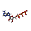
| #5: Chemical | ChemComp-ATP / Adenosine triphosphate Mass: 507.181 Da / Num. of mol.: 1 / Source method: obtained synthetically / Formula: C10H16N5O13P3 / Comment: ATP, energy-carrying molecule*YM Mass: 507.181 Da / Num. of mol.: 1 / Source method: obtained synthetically / Formula: C10H16N5O13P3 / Comment: ATP, energy-carrying molecule*YM |
| #6: Chemical | ChemComp-CA /  Mass: 40.078 Da / Num. of mol.: 1 / Source method: obtained synthetically / Formula: Ca Mass: 40.078 Da / Num. of mol.: 1 / Source method: obtained synthetically / Formula: Ca |
| #7: Water | ChemComp-HOH / WaterMass: 18.015 Da / Num. of mol.: 427 / Source method: isolated from a natural source / Formula: H2O |
-Details
| Nonpolymer details | AN ORDERED MAGNESIUM ION IS OBSERVED BOUND TO ATP. |
|---|
-Experimental details
-Experiment
| Experiment | Method: X-RAY DIFFRACTION / Number of used crystals: 1 |
|---|
- Sample preparation
| Crystal | Density Matthews: 2.87 Å3/Da / Density % sol: 50.5 % | ||||||||||||||||||||||||||||||||||||||||||||||||||||||||||||||||||||||
|---|---|---|---|---|---|---|---|---|---|---|---|---|---|---|---|---|---|---|---|---|---|---|---|---|---|---|---|---|---|---|---|---|---|---|---|---|---|---|---|---|---|---|---|---|---|---|---|---|---|---|---|---|---|---|---|---|---|---|---|---|---|---|---|---|---|---|---|---|---|---|---|
| Crystal grow | pH: 7.5 / Details: pH 7.50 | ||||||||||||||||||||||||||||||||||||||||||||||||||||||||||||||||||||||
| Crystal grow | *PLUS Method: vapor diffusion, hanging drop | ||||||||||||||||||||||||||||||||||||||||||||||||||||||||||||||||||||||
| Components of the solutions | *PLUS
|
-Data collection
| Diffraction | Mean temperature: 93 K |
|---|---|
| Diffraction source | Source: SYNCHROTRON / Site: NSLS  / Beamline: X9B / Wavelength: 0.97946 / Beamline: X9B / Wavelength: 0.97946 |
| Detector | Type: MARRESEARCH / Detector: IMAGE PLATE / Date: Jun 15, 1998 |
| Radiation | Protocol: SINGLE WAVELENGTH / Monochromatic (M) / Laue (L): M / Scattering type: x-ray |
| Radiation wavelength | Wavelength: 0.97946 Å / Relative weight: 1 |
| Reflection | Resolution: 1.9→20 Å / Num. obs: 49913 / % possible obs: 100 % / Observed criterion σ(I): 0 / Redundancy: 3.8 % / Rmerge(I) obs: 0.081 |
| Reflection shell | Resolution: 1.9→1.93 Å / Redundancy: 3.5 % / Rmerge(I) obs: 0.36 / % possible all: 99 |
| Reflection | *PLUS Redundancy: 3.77 % |
| Reflection shell | *PLUS % possible obs: 99.9 % |
- Processing
| Software |
| ||||||||||||||||||||||||||||||||||||||||||||||||||||||||||||
|---|---|---|---|---|---|---|---|---|---|---|---|---|---|---|---|---|---|---|---|---|---|---|---|---|---|---|---|---|---|---|---|---|---|---|---|---|---|---|---|---|---|---|---|---|---|---|---|---|---|---|---|---|---|---|---|---|---|---|---|---|---|
| Refinement | Method to determine structure: MOLECULAR REPLACEMENT Starting model: RABBIT SKELETAL MUSCLE ACTIN-HUMAN GELSOLIN SEGMENT 1 COMPLEX Resolution: 1.9→20 Å / Cross valid method: THROUGHOUT / σ(F): 0
| ||||||||||||||||||||||||||||||||||||||||||||||||||||||||||||
| Displacement parameters | Biso mean: 23.71 Å2 | ||||||||||||||||||||||||||||||||||||||||||||||||||||||||||||
| Refinement step | Cycle: LAST / Resolution: 1.9→20 Å
| ||||||||||||||||||||||||||||||||||||||||||||||||||||||||||||
| Refine LS restraints |
| ||||||||||||||||||||||||||||||||||||||||||||||||||||||||||||
| Xplor file |
| ||||||||||||||||||||||||||||||||||||||||||||||||||||||||||||
| Software | *PLUS Name: X-PLOR / Version: 3.851 / Classification: refinement | ||||||||||||||||||||||||||||||||||||||||||||||||||||||||||||
| Refinement | *PLUS Rfactor obs: 0.1915 / Rfactor Rfree: 0.2304 / Rfactor Rwork: 0.1915 | ||||||||||||||||||||||||||||||||||||||||||||||||||||||||||||
| Solvent computation | *PLUS | ||||||||||||||||||||||||||||||||||||||||||||||||||||||||||||
| Displacement parameters | *PLUS | ||||||||||||||||||||||||||||||||||||||||||||||||||||||||||||
| Refine LS restraints | *PLUS
|
