 Movie
Movie Controller


+ Open data

- Basic information
| Entry | Database: PDB / ID: 1wos | ||||||
|---|---|---|---|---|---|---|---|
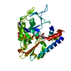
| Title | Crystal Structure of T-protein of the Glycine Cleavage System | ||||||
 Components Components | Aminomethyltransferase | ||||||
 Keywords Keywords | TRANSFERASE / aminomethyltransferase / T-protein | ||||||
| Function / homology |  Function and homology informationaminomethyltransferase / aminomethyltransferase activity / glycine decarboxylation via glycine cleavage system / transaminase activity / cytosol Function and homology informationaminomethyltransferase / aminomethyltransferase activity / glycine decarboxylation via glycine cleavage system / transaminase activity / cytosolSimilarity search - Function | ||||||
| Biological species |   Thermotoga maritima (bacteria) Thermotoga maritima (bacteria) | ||||||
| Method | X-RAY DIFFRACTION / SYNCHROTRON / Crystal 1 SINGLE WAVELENGTH PROTOCOL, Crysatl 2 MAD PROTOCOL / Resolution: 1.84 Å | ||||||
 Authors Authors | Lee, H.H. / Kim, D.J. / Ahn, H.J. / Ha, J.Y. / Suh, S.W. | ||||||
 Citation Citation | Journal: J.Biol.Chem. / Year: 2004 Title: Crystal Structure of T-protein of the Glycine Cleavage System: Cofactor binding, insights into H-protein recognition, and molecular basis for understanding nonketotic hyperglycinemia Authors: Lee, H.H. / Kim, D.J. / Ahn, H.J. / Ha, J.Y. / Suh, S.W. | ||||||
| History |
|
- Structure visualization
| Structure viewer | Molecule: MolmilJmol/JSmol |
|---|
- Downloads & links
-Download
| PDBx/mmCIF format | 1wos.cif.gz | 87 KB | Display | PDBx/mmCIF format |
|---|---|---|---|---|
| PDB format | pdb1wos.ent.gz | 65.1 KB | Display | PDB format |
| PDBx/mmJSON format | 1wos.json.gz | Tree view | PDBx/mmJSON format | |
| Others |  Other downloads Other downloads |
-Validation report
| Arichive directory | https://data.pdbj.org/pub/pdb/validation_reports/wo/1wosftp://data.pdbj.org/pub/pdb/validation_reports/wo/1wos | HTTPS FTP |
|---|
-Related structure data


-Links
 PDBj
PDBj- Assembly
| Deposited unit | 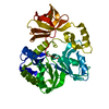
| ||||||||
|---|---|---|---|---|---|---|---|---|---|
| 1 |
| ||||||||
| Unit cell |
|
-Components
| #1: Protein | / Glycine cleavage system T protein Mass: 40381.445 Da / Num. of mol.: 1 Source method: isolated from a genetically manipulated source Source: (gene. exp.) Thermotoga maritima (bacteria) / Plasmid: pET21a / Production host: Escherichia coli (E. coli) / Strain (production host): B834(DE3) / References: UniProt: Q9WY54, aminomethyltransferase |
|---|---|
| #2: Water | ChemComp-HOH / Water Mass: 18.015 Da / Num. of mol.: 277 / Source method: isolated from a natural source / Formula: H2O Mass: 18.015 Da / Num. of mol.: 277 / Source method: isolated from a natural source / Formula: H2O |
-Experimental details
-Experiment
| Experiment | Method: X-RAY DIFFRACTION / Number of used crystals: 2 |
|---|
- Sample preparation
| Crystal | Density Matthews: 2.607 Å3/Da / Density % sol: 53 % |
|---|---|
| Crystal grow | Temperature: 297 K / Method: vapor diffusion, hanging drop / pH: 4.25 Details: PEG 3350, sodium dihydrogen phosphate, pH 4.25, VAPOR DIFFUSION, HANGING DROP, temperature 297K |
-Data collection
| Diffraction |
| |||||||||||||||
|---|---|---|---|---|---|---|---|---|---|---|---|---|---|---|---|---|
| Diffraction source | Source: SYNCHROTRON / Site: Photon Factory  / Beamline: BL-18B / Wavelength: 0.98020, 0.97947, 0.97935, 0.9500 / Beamline: BL-18B / Wavelength: 0.98020, 0.97947, 0.97935, 0.9500 | |||||||||||||||
| Detector | Type: ADSC QUANTUM 4 / Detector: CCD / Date: Feb 23, 2004 / Details: mirrors | |||||||||||||||
| Radiation | Monochromator: GRAPHITE / Protocol: MAD / Monochromatic (M) / Laue (L): M / Scattering type: x-ray | |||||||||||||||
| Radiation wavelength |
| |||||||||||||||
| Reflection | Resolution: 1.84→20 Å / Num. all: 38084 / Num. obs: 37551 / % possible obs: 98.6 % / Observed criterion σ(F): 0 / Observed criterion σ(I): 0 / Biso Wilson estimate: 11.3 Å2 | |||||||||||||||
| Reflection shell | Resolution: 1.84→1.91 Å / % possible all: 90 |
- Processing
| Software |
| ||||||||||||||||||||||||||||||||||||
|---|---|---|---|---|---|---|---|---|---|---|---|---|---|---|---|---|---|---|---|---|---|---|---|---|---|---|---|---|---|---|---|---|---|---|---|---|---|
| Refinement | Method to determine structure: Crystal 1 SINGLE WAVELENGTH PROTOCOL, Crysatl 2 MAD PROTOCOL Resolution: 1.84→19.99 Å / Rfactor Rfree error: 0.004 / Data cutoff high absF: 281265.65 / Data cutoff low absF: 0 / Isotropic thermal model: RESTRAINED / Cross valid method: THROUGHOUT / σ(F): 0 / Stereochemistry target values: Engh & Huber
| ||||||||||||||||||||||||||||||||||||
| Solvent computation | Solvent model: FLAT MODEL / Bsol: 45.8796 Å2 / ksol: 0.373529 e/Å3 | ||||||||||||||||||||||||||||||||||||
| Displacement parameters | Biso mean: 19.1 Å2
| ||||||||||||||||||||||||||||||||||||
| Refine analyze |
| ||||||||||||||||||||||||||||||||||||
| Refinement step | Cycle: LAST / Resolution: 1.84→19.99 Å
| ||||||||||||||||||||||||||||||||||||
| Refine LS restraints |
| ||||||||||||||||||||||||||||||||||||
| LS refinement shell | Resolution: 1.84→1.96 Å / Rfactor Rfree error: 0.013 / Total num. of bins used: 6
| ||||||||||||||||||||||||||||||||||||
| Xplor file |
|
