 Movie
Movie Controller

[English] 日本語
 Yorodumi
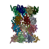
- PDB-1ryp: CRYSTAL STRUCTURE OF THE 20S PROTEASOME FROM YEAST AT 2.4 ANGSTRO... -
+ Open data

- Basic information
| Entry | Database: PDB / ID: 1ryp | ||||||
|---|---|---|---|---|---|---|---|
| Title | CRYSTAL STRUCTURE OF THE 20S PROTEASOME FROM YEAST AT 2.4 ANGSTROMS RESOLUTION | ||||||
 Components Components | (20S PROTEASOME Proteasome) x 14 Proteasome) x 14 | ||||||
 Keywords Keywords | MULTICATALYTIC PROTEINASE / 20S PROTEASOME / PROTEIN DEGRADATION / ANTIGEN PROCESSING / HYDROLASE / PROTEASE | ||||||
| Function / homology |  Function and homology information Function and homology informationproteasome core complex assembly / nuclear outer membrane-endoplasmic reticulum membrane network / Cross-presentation of soluble exogenous antigens (endosomes) / TNFR2 non-canonical NF-kB pathway / proteasomal ubiquitin-independent protein catabolic process / Ub-specific processing proteases / proteasome storage granule / endopeptidase activator activity / proteasome assembly / proteasome endopeptidase complex ...proteasome core complex assembly / nuclear outer membrane-endoplasmic reticulum membrane network / Cross-presentation of soluble exogenous antigens (endosomes) / TNFR2 non-canonical NF-kB pathway / proteasomal ubiquitin-independent protein catabolic process / Ub-specific processing proteases / proteasome storage granule / endopeptidase activator activity / proteasome assembly / proteasome endopeptidase complex / proteasome core complex, beta-subunit complex / proteasome core complex, alpha-subunit complex / threonine-type endopeptidase activity / Neutrophil degranulation / proteasome complex / proteasomal protein catabolic process / proteasome-mediated ubiquitin-dependent protein catabolic process / endopeptidase activity / mRNA binding / endoplasmic reticulum membrane / mitochondrion / nucleus / cytosol / cytoplasmSimilarity search - Function | ||||||
| Biological species |  Saccharomyces cerevisiae (brewer's yeast) Saccharomyces cerevisiae (brewer's yeast) | ||||||
| Method | X-RAY DIFFRACTION / SYNCHROTRON / Resolution: 1.9 Å | ||||||
 Authors Authors | Groll, M. / Ditzel, L. / Loewe, J. / Stock, D. / Bochtler, M. / Bartunik, H.D. / Huber, R. | ||||||
 Citation Citation | Journal: Nature / Year: 1997 Title: Structure of 20S proteasome from yeast at 2.4 A resolution. Authors: Groll, M. / Ditzel, L. / Lowe, J. / Stock, D. / Bochtler, M. / Bartunik, H.D. / Huber, R. | ||||||
| History |
|
- Structure visualization
| Structure viewer | Molecule: MolmilJmol/JSmol |
|---|
- Downloads & links
-Download
| PDBx/mmCIF format | 1ryp.cif.gz | 1.3 MB | Display | PDBx/mmCIF format |
|---|---|---|---|---|
| PDB format | pdb1ryp.ent.gz | 1 MB | Display | PDB format |
| PDBx/mmJSON format | 1ryp.json.gz | Tree view | PDBx/mmJSON format | |
| Others |  Other downloads Other downloads |
-Validation report
| Arichive directory | https://data.pdbj.org/pub/pdb/validation_reports/ry/1rypftp://data.pdbj.org/pub/pdb/validation_reports/ry/1ryp | HTTPS FTP |
|---|
-Related structure data
| Related structure data | 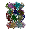 1pmaS S: Starting model for refinement |
|---|---|
| Similar structure data |


-Links
 PDBj
PDBj


- Assembly
| Deposited unit | 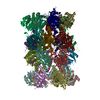
| ||||||||
|---|---|---|---|---|---|---|---|---|---|
| 1 |
| ||||||||
| Unit cell |
|
-Components
-Protein , 14 types, 28 molecules AOBPCQDRESFTGUHVIWJXKYLZM1N2
| #1: Protein | Proteasome Mass: 27316.037 Da / Num. of mol.: 2 / Mutation: CHAINS H, V, T1A, CHAIN L, Z, K33R / Source method: isolated from a natural source / Source: (natural) Saccharomyces cerevisiae (brewer's yeast) / References: UniProt: P21243, EC: 3.4.99.46#2: Protein | ProteasomeMass: 27191.828 Da / Num. of mol.: 2 / Mutation: CHAINS H, V, T1A, CHAIN L, Z, K33R / Source method: isolated from a natural source / Source: (natural) Saccharomyces cerevisiae (brewer's yeast) / References: UniProt: P23639, EC: 3.4.99.46#3: Protein | ProteasomeMass: 27050.416 Da / Num. of mol.: 2 / Mutation: CHAINS H, V, T1A, CHAIN L, Z, K33R / Source method: isolated from a natural source / Source: (natural) Saccharomyces cerevisiae (brewer's yeast) / References: UniProt: P23638, EC: 3.4.99.46#4: Protein | ProteasomeMass: 26903.330 Da / Num. of mol.: 2 / Mutation: CHAINS H, V, T1A, CHAIN L, Z, K33R / Source method: isolated from a natural source / Source: (natural) Saccharomyces cerevisiae (brewer's yeast) / References: UniProt: P40303, EC: 3.4.99.46#5: Protein | ProteasomeMass: 26544.789 Da / Num. of mol.: 2 / Mutation: CHAINS H, V, T1A, CHAIN L, Z, K33R / Source method: isolated from a natural source / Source: (natural) Saccharomyces cerevisiae (brewer's yeast) / References: UniProt: P32379, EC: 3.4.99.46#6: Protein | ProteasomeMass: 25502.805 Da / Num. of mol.: 2 / Mutation: CHAINS H, V, T1A, CHAIN L, Z, K33R / Source method: isolated from a natural source / Source: (natural) Saccharomyces cerevisiae (brewer's yeast) / References: UniProt: P40302, EC: 3.4.99.46#7: Protein | ProteasomeMass: 26892.482 Da / Num. of mol.: 2 / Mutation: CHAINS H, V, T1A, CHAIN L, Z, K33R / Source method: isolated from a natural source / Source: (natural) Saccharomyces cerevisiae (brewer's yeast) / References: UniProt: P21242, EC: 3.4.99.46#8: Protein | ProteasomeMass: 22401.266 Da / Num. of mol.: 2 / Mutation: CHAINS H, V, T1A, CHAIN L, Z, K33R / Source method: isolated from a natural source / Source: (natural) Saccharomyces cerevisiae (brewer's yeast) / References: UniProt: P38624, EC: 3.4.99.46#9: Protein | ProteasomeMass: 23987.254 Da / Num. of mol.: 2 / Mutation: CHAINS H, V, T1A, CHAIN L, Z, K33R / Source method: isolated from a natural source / Source: (natural) Saccharomyces cerevisiae (brewer's yeast) / References: UniProt: P25043, EC: 3.4.99.46#10: Protein | ProteasomeMass: 22496.645 Da / Num. of mol.: 2 / Mutation: CHAINS H, V, T1A, CHAIN L, Z, K33R / Source method: isolated from a natural source / Source: (natural) Saccharomyces cerevisiae (brewer's yeast) / References: UniProt: P25451, EC: 3.4.99.46#11: Protein | ProteasomeMass: 22545.676 Da / Num. of mol.: 2 / Mutation: CHAINS H, V, T1A, CHAIN L, Z, K33R / Source method: isolated from a natural source / Source: (natural) Saccharomyces cerevisiae (brewer's yeast) / References: UniProt: P22141, EC: 3.4.99.46#12: Protein | ProteasomeMass: 23353.262 Da / Num. of mol.: 2 / Mutation: CHAINS H, V, T1A, CHAIN L, Z, K33R / Source method: isolated from a natural source / Source: (natural) Saccharomyces cerevisiae (brewer's yeast) / References: UniProt: P30656, EC: 3.4.99.46#13: Protein | ProteasomeMass: 24883.928 Da / Num. of mol.: 2 / Mutation: CHAINS H, V, T1A, CHAIN L, Z, K33R / Source method: isolated from a natural source / Source: (natural) Saccharomyces cerevisiae (brewer's yeast) / References: UniProt: P23724, EC: 3.4.99.46#14: Protein | ProteasomeMass: 25945.496 Da / Num. of mol.: 2 / Mutation: CHAINS H, V, T1A, CHAIN L, Z, K33R / Source method: isolated from a natural source / Source: (natural) Saccharomyces cerevisiae (brewer's yeast) / References: UniProt: P30657, EC: 3.4.99.46 |
|---|
-Non-polymers , 2 types, 2928 molecules 

| #15: Chemical | ChemComp-MG /  Mass: 24.305 Da / Num. of mol.: 20 / Source method: obtained synthetically / Formula: Mg Mass: 24.305 Da / Num. of mol.: 20 / Source method: obtained synthetically / Formula: Mg#16: Water | ChemComp-HOH / | WaterMass: 18.015 Da / Num. of mol.: 2908 / Source method: isolated from a natural source / Formula: H2O |
|---|
-Experimental details
-Experiment
| Experiment | Method: X-RAY DIFFRACTION / Number of used crystals: 1 |
|---|
- Sample preparation
| Crystal | Density Matthews: 3.53 Å3/Da / Density % sol: 65.2 % | ||||||||||||||||||||||||||||||||||||||||||||||||||
|---|---|---|---|---|---|---|---|---|---|---|---|---|---|---|---|---|---|---|---|---|---|---|---|---|---|---|---|---|---|---|---|---|---|---|---|---|---|---|---|---|---|---|---|---|---|---|---|---|---|---|---|
| Crystal grow | pH: 6.9 / Details: pH 6.9 | ||||||||||||||||||||||||||||||||||||||||||||||||||
| Crystal grow | *PLUS Temperature: 24 ℃ / Method: vapor diffusion, hanging drop | ||||||||||||||||||||||||||||||||||||||||||||||||||
| Components of the solutions | *PLUS
|
-Data collection
| Diffraction | Mean temperature: 90 K |
|---|---|
| Diffraction source | Source: SYNCHROTRON / Site: MPG/DESY, HAMBURG  / Beamline: BW6 / Wavelength: 1.1 / Beamline: BW6 / Wavelength: 1.1 |
| Detector | Type: MARRESEARCH / Detector: IMAGE PLATE / Date: Jun 1, 1997 |
| Radiation | Monochromatic (M) / Laue (L): M / Scattering type: x-ray |
| Radiation wavelength | Wavelength: 1.1 Å / Relative weight: 1 |
| Reflection | Resolution: 1.9→50 Å / Num. obs: 778118 / % possible obs: 93.6 % / Observed criterion σ(I): 2 / Redundancy: 2.9 % / Rmerge(I) obs: 0.081 / Net I/σ(I): 2 |
| Reflection shell | Highest resolution: 1.97 Å / Redundancy: 1.8 % / Rmerge(I) obs: 0.487 / Mean I/σ(I) obs: 2 / % possible all: 93.6 |
- Processing
| Software |
| ||||||||||||||||||||||||||||||||||||||||||||||||||||||||||||||||||||||||||||||||
|---|---|---|---|---|---|---|---|---|---|---|---|---|---|---|---|---|---|---|---|---|---|---|---|---|---|---|---|---|---|---|---|---|---|---|---|---|---|---|---|---|---|---|---|---|---|---|---|---|---|---|---|---|---|---|---|---|---|---|---|---|---|---|---|---|---|---|---|---|---|---|---|---|---|---|---|---|---|---|---|---|---|
| Refinement | Starting model: PDB ENTRY 1PMA Resolution: 1.9→50 Å / Rfactor Rfree error: 0 / Data cutoff high absF: 100000000 / Data cutoff low absF: 0.1 / Isotropic thermal model: RESTRAINED / σ(F): 2
| ||||||||||||||||||||||||||||||||||||||||||||||||||||||||||||||||||||||||||||||||
| Displacement parameters | Biso mean: 36.4 Å2
| ||||||||||||||||||||||||||||||||||||||||||||||||||||||||||||||||||||||||||||||||
| Refinement step | Cycle: LAST / Resolution: 1.9→50 Å
| ||||||||||||||||||||||||||||||||||||||||||||||||||||||||||||||||||||||||||||||||
| Refine LS restraints |
| ||||||||||||||||||||||||||||||||||||||||||||||||||||||||||||||||||||||||||||||||
| Refine LS restraints NCS | Rms dev Biso : 2 Å2 / Rms dev position: 0.15 Å / Weight Biso : 2 | ||||||||||||||||||||||||||||||||||||||||||||||||||||||||||||||||||||||||||||||||
| Software | *PLUS Name: X-PLOR / Version: 3.1 / Classification: refinement | ||||||||||||||||||||||||||||||||||||||||||||||||||||||||||||||||||||||||||||||||
| Refinement | *PLUS Rfactor Rfree: 0.33 | ||||||||||||||||||||||||||||||||||||||||||||||||||||||||||||||||||||||||||||||||
| Solvent computation | *PLUS | ||||||||||||||||||||||||||||||||||||||||||||||||||||||||||||||||||||||||||||||||
| Displacement parameters | *PLUS | ||||||||||||||||||||||||||||||||||||||||||||||||||||||||||||||||||||||||||||||||
| Refine LS restraints | *PLUS
|
