Resolution: 1.9→1.97 Å / Redundancy: 2.9 % / Mean I/σ(I) obs: 4.9 / Num. unique all: 17439 / Rsym value: 0.202 / % possible all: 69.1
Reflection
*PLUS
Lowest resolution: 30 Å / Rmerge(I) obs: 0.055
Reflection shell
*PLUS
Highest resolution: 1.9 Å / % possible obs: 69.1 % / Rmerge(I) obs: 0.236
-
Processing
Software
Name
Version
Classification
CNS
1.1
refinement
DENZO
datareduction
SCALEPACK
datascaling
SHARP
phasing
Refinement
Method to determine structure: MIR / Resolution: 2→29.55 Å / Rfactor Rfree error: 0.002 / Isotropic thermal model: RESTRAINED / Cross valid method: THROUGHOUT / σ(F): 0 / Stereochemistry target values: Engh & Huber Details: maximum likelihood target using amplitudes. WATERS 1-2 HAVE COORDINATION SPHERES REMINISCENT OF CATIONS, BUT COULD NOT BE DEFINITIVELY IDENTIFIED AS IONS BECAUSE THEIR B-FACTORS ARE NOT ...Details: maximum likelihood target using amplitudes. WATERS 1-2 HAVE COORDINATION SPHERES REMINISCENT OF CATIONS, BUT COULD NOT BE DEFINITIVELY IDENTIFIED AS IONS BECAUSE THEIR B-FACTORS ARE NOT UNREASONABLY LOW COMPARED TO THE SURROUNDING ATOMS, AND THERE ARE NO CORRESPONDING PEAKS IN THE ANOMALOUS DIFFERENCE DENSITY
In the structure databanks used in Yorodumi, some data are registered as the other names, "COVID-19 virus" and "2019-nCoV". Here are the details of the virus and the list of structure data.
Jan 31, 2019. EMDB accession codes are about to change! (news from PDBe EMDB page)
EMDB accession codes are about to change! (news from PDBe EMDB page)
The allocation of 4 digits for EMDB accession codes will soon come to an end. Whilst these codes will remain in use, new EMDB accession codes will include an additional digit and will expand incrementally as the available range of codes is exhausted. The current 4-digit format prefixed with “EMD-” (i.e. EMD-XXXX) will advance to a 5-digit format (i.e. EMD-XXXXX), and so on. It is currently estimated that the 4-digit codes will be depleted around Spring 2019, at which point the 5-digit format will come into force.
The EM Navigator/Yorodumi systems omit the EMD- prefix.
Related info.:Q: What is EMD? / ID/Accession-code notation in Yorodumi/EM Navigator
Yorodumi is a browser for structure data from EMDB, PDB, SASBDB, etc.
This page is also the successor to EM Navigator detail page, and also detail information page/front-end page for Omokage search.
The word "yorodu" (or yorozu) is an old Japanese word meaning "ten thousand". "mi" (miru) is to see.
Related info.:EMDB / PDB / SASBDB / Comparison of 3 databanks / Yorodumi Search / Aug 31, 2016. New EM Navigator & Yorodumi / Yorodumi Papers / Jmol/JSmol / Function and homology information / Changes in new EM Navigator and Yorodumi
 Movie
Movie Controller

 Yorodumi
 Open data
 Basic information
 Components
 Keywords
 TRANSFERASE / carboxyl transferase / domain duplication / multienzyme complex / transcarboxylase
 Function and homology information
 Authors
 Citation
 Structure visualization
 Downloads & links
 Other downloads
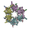


 PDBj
PDBj Assembly


 Mass: 112.411 Da / Num. of mol.: 3 / Source method: obtained synthetically / Formula: Cd
 Mass: 867.607 Da / Num. of mol.: 6 / Source method: obtained synthetically / Formula: C25H40N7O19P3S
 Mass: 118.174 Da / Num. of mol.: 1 / Source method: obtained synthetically / Formula: C6H14O2 / Comment: precipitant*YM
 Mass: 18.015 Da / Num. of mol.: 3390 / Source method: isolated from a natural source / Formula: H2O
 Sample preparation
 / Beamline: 19-ID / Wavelength: 1 Å
 Processing