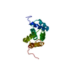
 Movie
Movie Controller


+ Open data

- Basic information
| Entry | Database: PDB / ID: 1lsu | ||||||
|---|---|---|---|---|---|---|---|
| Title | KTN Bsu222 Crystal Structure in Complex with NADH | ||||||
 Components Components | Conserved hypothetical protein yuaA | ||||||
 Keywords Keywords |  TRANSPORT PROTEIN / KTN domain / NAD / RCK domain / Potassium Transport / Potassium Channel / KtrA / Rossmann Fold TRANSPORT PROTEIN / KTN domain / NAD / RCK domain / Potassium Transport / Potassium Channel / KtrA / Rossmann Fold | ||||||
| Function / homology |  Function and homology information Function and homology informationmonoatomic cation transmembrane transporter activity / potassium ion transport / identical protein binding / plasma membraneSimilarity search - Function | ||||||
| Biological species |  Bacillus subtilis (bacteria) Bacillus subtilis (bacteria) | ||||||
| Method | X-RAY DIFFRACTION / SYNCHROTRON / MAD, MIR / Resolution: 2.85 Å | ||||||
 Authors Authors | Roosild, T.P. / Miller, S. / Booth, I.R. / Choe, S. | ||||||
 Citation Citation | Journal: Cell(Cambridge,Mass.) / Year: 2002 Title: A mechanism of regulating transmembrane potassium flux through a ligand-mediated conformational switch. Authors: Roosild, T.P. / Miller, S. / Booth, I.R. / Choe, S. | ||||||
| History |
| ||||||
| Remark 999 | SEQUENCE Authors state the sequence present in the B. subtilis lab strain from which they cloned ...SEQUENCE Authors state the sequence present in the B. subtilis lab strain from which they cloned may be a strain type polymorphism, or an error in the database itself. |
- Structure visualization
| Structure viewer | Molecule: MolmilJmol/JSmol |
|---|
- Downloads & links
-Download
| PDBx/mmCIF format | 1lsu.cif.gz | 66 KB | Display | PDBx/mmCIF format |
|---|---|---|---|---|
| PDB format | pdb1lsu.ent.gz | 50.1 KB | Display | PDB format |
| PDBx/mmJSON format | 1lsu.json.gz | Tree view | PDBx/mmJSON format | |
| Others |  Other downloads Other downloads |
-Validation report
| Arichive directory | https://data.pdbj.org/pub/pdb/validation_reports/ls/1lsuftp://data.pdbj.org/pub/pdb/validation_reports/ls/1lsu | HTTPS FTP |
|---|
-Related structure data


-Links
 PDBj
PDBj


- Assembly
| Deposited unit | 
| ||||||||
|---|---|---|---|---|---|---|---|---|---|
| 1 | 
| ||||||||
| 2 | 
| ||||||||
| Unit cell |
| ||||||||
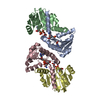
| Details | The KTN biological unit consists of two helix-swapped monomers. The asymmetric unit contains one biological unit formed by chains A & B |
-Components
| #1: Protein | Mass: 16225.580 Da / Num. of mol.: 2 / Fragment: KTN Domain, Residues 1-143 / Mutation: C22V Source method: isolated from a genetically manipulated source Source: (gene. exp.) Bacillus subtilis (bacteria) / Gene: KtrA / Plasmid: pHis8 / Species (production host): Escherichia coli / Production host: Escherichia coli BL21 (bacteria) / Strain (production host): BL-21 / References: UniProt: O32080#2: Chemical | Nicotinamide adenine dinucleotide  Mass: 665.441 Da / Num. of mol.: 2 / Source method: obtained synthetically / Formula: C21H29N7O14P2 Mass: 665.441 Da / Num. of mol.: 2 / Source method: obtained synthetically / Formula: C21H29N7O14P2 |
|---|
-Experimental details
-Experiment
| Experiment | Method: X-RAY DIFFRACTION / Number of used crystals: 2 |
|---|
- Sample preparation
| Crystal | Density Matthews: 3.05 Å3/Da / Density % sol: 59.62 % | ||||||||||||||||||||||||||||||||||||||||||
|---|---|---|---|---|---|---|---|---|---|---|---|---|---|---|---|---|---|---|---|---|---|---|---|---|---|---|---|---|---|---|---|---|---|---|---|---|---|---|---|---|---|---|---|
| Crystal grow | Temperature: 298 K / Method: vapor diffusion, hanging drop / pH: 6 Details: Ammonium Sulfate, pH 6, VAPOR DIFFUSION, HANGING DROP, temperature 298K | ||||||||||||||||||||||||||||||||||||||||||
| Crystal grow | *PLUS pH: 8 | ||||||||||||||||||||||||||||||||||||||||||
| Components of the solutions | *PLUS
|
-Data collection
| Diffraction |
| |||||||||||||||||||||
|---|---|---|---|---|---|---|---|---|---|---|---|---|---|---|---|---|---|---|---|---|---|---|
| Diffraction source |
| |||||||||||||||||||||
| Detector | Type: ADSC QUANTUM 4 / Detector: CCD / Date: Jun 4, 2001 | |||||||||||||||||||||
| Radiation |
| |||||||||||||||||||||
| Radiation wavelength |
| |||||||||||||||||||||
| Reflection | Resolution: 2.85→30 Å / Num. all: 9667 / Num. obs: 9635 / % possible obs: 99.6 % / Observed criterion σ(F): 2 / Observed criterion σ(I): 2 / Biso Wilson estimate: 71.1 Å2 / Rmerge(I) obs: 0.065 / Net I/σ(I): 16.4 | |||||||||||||||||||||
| Reflection shell | Resolution: 2.85→2.95 Å / Rmerge(I) obs: 0.343 / Mean I/σ(I) obs: 3.5 / Num. unique all: 947 / % possible all: 100 | |||||||||||||||||||||
| Reflection | *PLUS % possible obs: 100 % / Rmerge(I) obs: 0.07 | |||||||||||||||||||||
| Reflection shell | *PLUS Rmerge(I) obs: 0.343 |

- Processing
| Software |
| ||||||||||||||||||||||||||||||||||||||||||||||||||||||||||||||||||||||||||||||||
|---|---|---|---|---|---|---|---|---|---|---|---|---|---|---|---|---|---|---|---|---|---|---|---|---|---|---|---|---|---|---|---|---|---|---|---|---|---|---|---|---|---|---|---|---|---|---|---|---|---|---|---|---|---|---|---|---|---|---|---|---|---|---|---|---|---|---|---|---|---|---|---|---|---|---|---|---|---|---|---|---|---|
| Refinement | Method to determine structure: MAD, MIR / Resolution: 2.85→30 Å / Rfactor Rfree error: 0.013 / Data cutoff high absF: 1669970.48 / Data cutoff high rms absF: 1669970.48 / Data cutoff low absF: 0 / Isotropic thermal model: RESTRAINED / Cross valid method: THROUGHOUT / σ(F): 0 / σ(I): 0 / Stereochemistry target values: Engh & Huber
| ||||||||||||||||||||||||||||||||||||||||||||||||||||||||||||||||||||||||||||||||
| Solvent computation | Solvent model: FLAT MODEL / Bsol: 41.8566 Å2 / ksol: 0.354957 e/Å3 | ||||||||||||||||||||||||||||||||||||||||||||||||||||||||||||||||||||||||||||||||
| Displacement parameters | Biso mean: 70.4 Å2
| ||||||||||||||||||||||||||||||||||||||||||||||||||||||||||||||||||||||||||||||||
| Refine analyze |
| ||||||||||||||||||||||||||||||||||||||||||||||||||||||||||||||||||||||||||||||||
| Refinement step | Cycle: LAST / Resolution: 2.85→30 Å
| ||||||||||||||||||||||||||||||||||||||||||||||||||||||||||||||||||||||||||||||||
| Refine LS restraints |
| ||||||||||||||||||||||||||||||||||||||||||||||||||||||||||||||||||||||||||||||||
| LS refinement shell | Resolution: 2.85→3.03 Å / Rfactor Rfree error: 0.043 / Total num. of bins used: 6
| ||||||||||||||||||||||||||||||||||||||||||||||||||||||||||||||||||||||||||||||||
| Xplor file |
| ||||||||||||||||||||||||||||||||||||||||||||||||||||||||||||||||||||||||||||||||
| Refinement | *PLUS Lowest resolution: 30 Å / Rfactor Rfree: 0.329 / Rfactor Rwork: 0.279 | ||||||||||||||||||||||||||||||||||||||||||||||||||||||||||||||||||||||||||||||||
| Solvent computation | *PLUS | ||||||||||||||||||||||||||||||||||||||||||||||||||||||||||||||||||||||||||||||||
| Displacement parameters | *PLUS | ||||||||||||||||||||||||||||||||||||||||||||||||||||||||||||||||||||||||||||||||
| Refine LS restraints | *PLUS
| ||||||||||||||||||||||||||||||||||||||||||||||||||||||||||||||||||||||||||||||||
| LS refinement shell | *PLUS Rfactor Rfree: 0.467 / Rfactor Rwork: 0.43 |
