 Movie
Movie Controller


+ Open data

- Basic information
| Entry | Database: PDB / ID: 1lo0 | ||||||
|---|---|---|---|---|---|---|---|
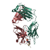
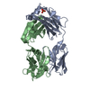
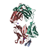
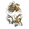
| Title | Catalytic Retro-Diels-Alderase Transition State Analogue Complex | ||||||
 Components Components |
| ||||||
 Keywords Keywords |  IMMUNE SYSTEM / catalytic antibody / Fab fragment IMMUNE SYSTEM / catalytic antibody / Fab fragment | ||||||
| Function / homology |  Function and homology information Function and homology informationpositive regulation of B cell activation / humoral immune response mediated by circulating immunoglobulin / early endosome to late endosome transport / phagocytosis, recognition / positive regulation of type IIa hypersensitivity / regulation of proteolysis / positive regulation of type I hypersensitivity / antibody-dependent cellular cytotoxicity / Fc-gamma receptor I complex binding / phagocytosis, engulfment ...positive regulation of B cell activation / humoral immune response mediated by circulating immunoglobulin / early endosome to late endosome transport / phagocytosis, recognition / positive regulation of type IIa hypersensitivity / regulation of proteolysis / positive regulation of type I hypersensitivity / antibody-dependent cellular cytotoxicity / Fc-gamma receptor I complex binding / phagocytosis, engulfment / IgG immunoglobulin complex / endosome to lysosome transport / positive regulation of endocytosis / immunoglobulin complex, circulating / immunoglobulin receptor binding / antigen processing and presentation / immunoglobulin mediated immune response / positive regulation of phagocytosis / complement activation, classical pathway / multivesicular body / antigen binding / B cell differentiation / response to bacterium / positive regulation of immune response / antibacterial humoral response / extracellular space / extracellular region / metal ion binding / plasma membrane / cytosolSimilarity search - Function | ||||||
| Biological species |  Mus musculus (house mouse) Mus musculus (house mouse) | ||||||
| Method | X-RAY DIFFRACTION / MOLECULAR REPLACEMENT / Resolution: 2 Å | ||||||
 Authors Authors | Hugot, M. / Reymond, J.L. / Baumann, U. | ||||||
 Citation Citation | Journal: Proc.Natl.Acad.Sci.USA / Year: 2002 Title: A structural basis for the activity of retro-Diels-Alder catalytic antibodies: evidence for a catalytic aromatic residue. Authors: Hugot, M. / Bensel, N. / Vogel, M. / Reymond, M.T. / Stadler, B. / Reymond, J.L. / Baumann, U. | ||||||
| History |
| ||||||
| Remark 999 | SEQUENCE At the time of processing, this sequence has not yet been deposited in a sequence database. |
- Structure visualization
| Structure viewer | Molecule: MolmilJmol/JSmol |
|---|
- Downloads & links
-Download
| PDBx/mmCIF format | 1lo0.cif.gz | 166.3 KB | Display | PDBx/mmCIF format |
|---|---|---|---|---|
| PDB format | pdb1lo0.ent.gz | 134.4 KB | Display | PDB format |
| PDBx/mmJSON format | 1lo0.json.gz | Tree view | PDBx/mmJSON format | |
| Others |  Other downloads Other downloads |
-Validation report
| Arichive directory | https://data.pdbj.org/pub/pdb/validation_reports/lo/1lo0ftp://data.pdbj.org/pub/pdb/validation_reports/lo/1lo0 | HTTPS FTP |
|---|
-Related structure data
| Related structure data |  1lo2C  1lo3C  1lo4C  28b4S S: Starting model for refinement C: citing same article ( |
|---|---|
| Similar structure data |


-Links
 PDBj
PDBj


- Assembly
| Deposited unit | 
| ||||||||
|---|---|---|---|---|---|---|---|---|---|
| 1 | 
| ||||||||
| 2 | 
| ||||||||
| Unit cell |
| ||||||||
| Noncrystallographic symmetry (NCS) | NCS domain: (Details: RESTRAINED) |
-Components
| #1: Antibody | Mass: 24268.992 Da / Num. of mol.: 2 / Fragment: Fab fragment / Source method: isolated from a natural source Details: The antibodies were isolated from hybridoma cells and Fab fragments were generated by papain digestion. Source: (natural) Mus musculus (house mouse) / References: UniProt: P01837, UniProt: A2NHM3*PLUS#2: Antibody | Mass: 23704.830 Da / Num. of mol.: 2 / Source method: isolated from a natural source Details: The antibodies were isolated from hybridoma cells and Fab fragments were generated by papain digestion. Source: (natural) Mus musculus (house mouse) / References: UniProt: P01865#3: Chemical |   Mass: 335.357 Da / Num. of mol.: 2 / Source method: obtained synthetically / Formula: C19H17N3O3 Mass: 335.357 Da / Num. of mol.: 2 / Source method: obtained synthetically / Formula: C19H17N3O3#4: Water | ChemComp-HOH / | Water Mass: 18.015 Da / Num. of mol.: 105 / Source method: isolated from a natural source / Formula: H2O Mass: 18.015 Da / Num. of mol.: 105 / Source method: isolated from a natural source / Formula: H2O |
|---|
-Experimental details
-Experiment
| Experiment | Method: X-RAY DIFFRACTION / Number of used crystals: 1 |
|---|
- Sample preparation
| Crystal | Density Matthews: 2.53 Å3/Da / Density % sol: 51.34 % | ||||||||||||||||||||||||||||||||||||
|---|---|---|---|---|---|---|---|---|---|---|---|---|---|---|---|---|---|---|---|---|---|---|---|---|---|---|---|---|---|---|---|---|---|---|---|---|---|
| Crystal grow | Temperature: 290 K / Method: vapor diffusion, hanging drop / pH: 7 Details: PEG, pH 7.0, VAPOR DIFFUSION, HANGING DROP, temperature 290K | ||||||||||||||||||||||||||||||||||||
| Crystal grow | *PLUS Temperature: 20 ℃ / pH: 7.8 / Method: vapor diffusion | ||||||||||||||||||||||||||||||||||||
| Components of the solutions | *PLUS
|
-Data collection
| Diffraction | Mean temperature: 110 K |
|---|---|
| Diffraction source | Source: ROTATING ANODE / Type: RIGAKU RU300 / Wavelength: 1.5418 Å |
| Detector | Type: RIGAKU RAXIS IV / Detector: IMAGE PLATE / Date: Jan 1, 2000 / Details: Yale mirrors |
| Radiation | Protocol: SINGLE WAVELENGTH / Monochromatic (M) / Laue (L): M / Scattering type: x-ray |
| Radiation wavelength | Wavelength: 1.5418 Å / Relative weight: 1 |
| Reflection | Resolution: 1.77→30.3 Å / Num. obs: 78457 / % possible obs: 95.9 % / Observed criterion σ(I): 0 / Biso Wilson estimate: 30 Å2 / Rmerge(I) obs: 0.051 / Rsym value: 0.051 / Net I/σ(I): 24.4 |
| Reflection shell | Resolution: 1.77→1.85 Å / Rmerge(I) obs: 0.136 / Mean I/σ(I) obs: 9.9 / Rsym value: 0.136 / % possible all: 0.959 |
| Reflection | *PLUS Num. obs: 74192 / % possible obs: 81.7 % / Num. measured all: 1025225 |
| Reflection shell | *PLUS % possible obs: 95.9 % |
- Processing
| Software |
| ||||||||||||||||||||||||||||||||||||||||||||||||||||||||||||
|---|---|---|---|---|---|---|---|---|---|---|---|---|---|---|---|---|---|---|---|---|---|---|---|---|---|---|---|---|---|---|---|---|---|---|---|---|---|---|---|---|---|---|---|---|---|---|---|---|---|---|---|---|---|---|---|---|---|---|---|---|---|
| Refinement | Method to determine structure: MOLECULAR REPLACEMENT Starting model: 28B4 Resolution: 2→30.3 Å / Isotropic thermal model: GROUP / Cross valid method: THROUGHOUT / σ(F): 0 / Stereochemistry target values: ENGH & HUBER Details: CRYSTALS ARE PSEUDO-MEROHEDRALLY TWINNED TWINNING LAW H,-K,-L TWINNING FRACTION 0.3787
| ||||||||||||||||||||||||||||||||||||||||||||||||||||||||||||
| Solvent computation | Solvent model: SOLVENT MASK / Bsol: 36.9 Å2 / ksol: 0.341 e/Å3 | ||||||||||||||||||||||||||||||||||||||||||||||||||||||||||||
| Displacement parameters | Biso mean: 25.3 Å2
| ||||||||||||||||||||||||||||||||||||||||||||||||||||||||||||
| Refine analyze |
| ||||||||||||||||||||||||||||||||||||||||||||||||||||||||||||
| Refinement step | Cycle: LAST / Resolution: 2→30.3 Å
| ||||||||||||||||||||||||||||||||||||||||||||||||||||||||||||
| Refine LS restraints |
| ||||||||||||||||||||||||||||||||||||||||||||||||||||||||||||
| LS refinement shell | Resolution: 2→2.07 Å / Total num. of bins used: 10
| ||||||||||||||||||||||||||||||||||||||||||||||||||||||||||||
| Refinement | *PLUS Rfactor Rfree: 0.256 / Rfactor Rwork: 0.204 | ||||||||||||||||||||||||||||||||||||||||||||||||||||||||||||
| Solvent computation | *PLUS | ||||||||||||||||||||||||||||||||||||||||||||||||||||||||||||
| Displacement parameters | *PLUS | ||||||||||||||||||||||||||||||||||||||||||||||||||||||||||||
| Refine LS restraints | *PLUS
|
