 Movie
Movie Controller


+ Open data

- Basic information
| Entry | Database: PDB / ID: 1kwa | ||||||
|---|---|---|---|---|---|---|---|
| Title | HUMAN CASK/LIN-2 PDZ DOMAIN | ||||||
 Components Components | HCASK/LIN-2 PROTEIN | ||||||
 Keywords Keywords |  KINASE / PDZ DOMAIN / NEUREXIN / SYNDECAN / RECEPTOR CLUSTERING KINASE / PDZ DOMAIN / NEUREXIN / SYNDECAN / RECEPTOR CLUSTERING | ||||||
| Function / homology |  Function and homology information Function and homology informationnegative regulation of cellular response to growth factor stimulus / guanylate kinase activity / Dopamine Neurotransmitter Release Cycle / neurexin family protein binding / regulation of neurotransmitter secretion / negative regulation of wound healing / nuclear lamina / calcium ion import / Assembly and cell surface presentation of NMDA receptors / Neurexins and neuroligins ...negative regulation of cellular response to growth factor stimulus / guanylate kinase activity / Dopamine Neurotransmitter Release Cycle / neurexin family protein binding / regulation of neurotransmitter secretion / negative regulation of wound healing / nuclear lamina / calcium ion import / Assembly and cell surface presentation of NMDA receptors / Neurexins and neuroligins / Sensory processing of sound by outer hair cells of the cochlea / Sensory processing of sound by inner hair cells of the cochlea / Nephrin family interactions / ciliary membrane / regulation of synaptic vesicle exocytosis / Syndecan interactions / negative regulation of cell-matrix adhesion / positive regulation of calcium ion import / basement membrane / negative regulation of keratinocyte proliferation / establishment of localization in cell / Schaffer collateral - CA1 synapse / nuclear matrix / cell-cell junction / actin cytoskeleton / presynaptic membrane / basolateral plasma membrane / vesicle / calmodulin binding / non-specific serine/threonine protein kinase / cell adhesion / phosphorylation / signaling receptor binding / focal adhesion / protein serine kinase activity / protein serine/threonine kinase activity / nucleolus / positive regulation of transcription by RNA polymerase II / ATP binding / plasma membrane / cytosol / cytoplasmSimilarity search - Function | ||||||
| Biological species |  Homo sapiens (human) Homo sapiens (human) | ||||||
| Method | X-RAY DIFFRACTION / SYNCHROTRON / MAD, SIR / Resolution: 1.93 Å | ||||||
 Authors Authors | Daniels, D.L. / Cohen, A.R. / Anderson, J.M. / Brunger, A.T. | ||||||
 Citation Citation | Journal: Nat.Struct.Biol. / Year: 1998 Title: Crystal structure of the hCASK PDZ domain reveals the structural basis of class II PDZ domain target recognition Authors: Daniels, D.L. / Cohen, A.R. / Anderson, J.M. / Brunger, A.T. #1: Journal: Cell(Cambridge,Mass.) / Year: 1996Title: Crystal Structures of a Complexed and Peptide-Free Membrane Protein-Binding Domain: Molecular Basis of Peptide Recognition by Pdz Authors: Doyle, D.A. / Lee, A. / Lewis, J. / Kim, E. / Sheng, M. / Mackinnon, R. | ||||||
| History |
|
- Structure visualization
| Structure viewer | Molecule: MolmilJmol/JSmol |
|---|
- Downloads & links
-Download
| PDBx/mmCIF format | 1kwa.cif.gz | 51.7 KB | Display | PDBx/mmCIF format |
|---|---|---|---|---|
| PDB format | pdb1kwa.ent.gz | 37.2 KB | Display | PDB format |
| PDBx/mmJSON format | 1kwa.json.gz | Tree view | PDBx/mmJSON format | |
| Others |  Other downloads Other downloads |
-Validation report
| Arichive directory | https://data.pdbj.org/pub/pdb/validation_reports/kw/1kwaftp://data.pdbj.org/pub/pdb/validation_reports/kw/1kwa | HTTPS FTP |
|---|
-Related structure data
| Similar structure data |
|---|


-Links
 PDBj
PDBj


- Assembly
| Deposited unit | 
| |||||||||||||||
|---|---|---|---|---|---|---|---|---|---|---|---|---|---|---|---|---|
| 1 | 
| |||||||||||||||
| 2 | 
| |||||||||||||||
| 3 | 
| |||||||||||||||
| 4 |
| |||||||||||||||
| 5 | 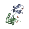
| |||||||||||||||
| Unit cell |
| |||||||||||||||
| Components on special symmetry positions |
| |||||||||||||||
| Noncrystallographic symmetry (NCS) | NCS oper: (Code: given Matrix: (0.417572, 0.546936, 0.7256), Vector : |
-Components
| #1: Protein | Mass: 10194.873 Da / Num. of mol.: 2 / Fragment: PDZ DOMAIN Source method: isolated from a genetically manipulated source Source: (gene. exp.) Homo sapiens (human) / Description: CLONED FROM A CDNA LIVER LIBRARY / Cellular location: MEMBRANE-ASSOCIATED / Gene: HCASK / Organ: LIVER / Plasmid: N-TERMINAL GST FUSION, AMP RESISTANT / Cellular location (production host): CYTOPLASM / Production host:  Escherichia coli (E. coli) / Strain (production host): DH5 ALPHA / References: UniProt: O14936 Escherichia coli (E. coli) / Strain (production host): DH5 ALPHA / References: UniProt: O14936#2: Chemical | Sulfate  Mass: 96.063 Da / Num. of mol.: 3 / Fragment: PDZ DOMAIN / Source method: obtained synthetically / Formula: SO4 Mass: 96.063 Da / Num. of mol.: 3 / Fragment: PDZ DOMAIN / Source method: obtained synthetically / Formula: SO4#3: Water | ChemComp-HOH / | Water Mass: 18.015 Da / Num. of mol.: 185 / Source method: isolated from a natural source / Formula: H2O Mass: 18.015 Da / Num. of mol.: 185 / Source method: isolated from a natural source / Formula: H2O |
|---|
-Experimental details
-Experiment
| Experiment | Method: X-RAY DIFFRACTION / Number of used crystals: 1 |
|---|
- Sample preparation
| Crystal | Density Matthews: 2.3 Å3/Da / Density % sol: 55 % | ||||||||||||||||||||||||||||||
|---|---|---|---|---|---|---|---|---|---|---|---|---|---|---|---|---|---|---|---|---|---|---|---|---|---|---|---|---|---|---|---|
| Crystal grow | Temperature: 294 K / pH: 6.5 Details: PROTEIN WAS CRYSTALLIZED IN 1.8M AMMONIUM SULFATE AND 100MM MES PH 6.5 AT 21 DEGREES C., temperature 294K | ||||||||||||||||||||||||||||||
| Crystal grow | *PLUS Temperature: 21 ℃ / Method: vapor diffusion, hanging drop | ||||||||||||||||||||||||||||||
| Components of the solutions | *PLUS
|
-Data collection
| Diffraction | Mean temperature: 110 K | |||||||||
|---|---|---|---|---|---|---|---|---|---|---|
| Diffraction source | Source: SYNCHROTRON / Site: SSRL  / Beamline: BL1-5 / Wavelength: 0.979 / Wavelength: 0.979, 1.033 / Beamline: BL1-5 / Wavelength: 0.979 / Wavelength: 0.979, 1.033 | |||||||||
| Detector | Type: MARRESEARCH / Detector: IMAGE PLATE AREA DETECTOR / Date: Dec 1, 1996 / Details: MIRROR | |||||||||
| Radiation | Monochromator: SI(111) / Monochromatic (M) / Laue (L): M / Scattering type: x-ray | |||||||||
| Radiation wavelength |
| |||||||||
| Reflection | Resolution: 1.93→30 Å / Num. obs: 14216 / % possible obs: 88.4 % / Observed criterion σ(I): 2 / Redundancy: 3.5 % / Biso Wilson estimate: 13 Å2 / Rsym value: 0.032 / Net I/σ(I): 21.6 | |||||||||
| Reflection shell | Resolution: 1.93→1.96 Å / Redundancy: 2 % / Mean I/σ(I) obs: 3.1 / Rsym value: 0.293 / % possible all: 78.1 | |||||||||
| Reflection | *PLUS Num. measured all: 50697 / Rmerge(I) obs: 0.032 | |||||||||
| Reflection shell | *PLUS % possible obs: 78.1 % / Rmerge(I) obs: 0.293 |
- Processing
| Refinement | Method to determine structure: MAD, SIR / Resolution: 1.93→30 Å / Rfactor Rfree error: 0.008 / Isotropic thermal model: RESTRAINED / Cross valid method: THROUGHOUT / σ(F): 0 / Details: BULK SOLVENT MODEL USED
| ||||||||||||||||||||||||||||||||||||||||||||||||||||||||||||||||||||||||||||||||
|---|---|---|---|---|---|---|---|---|---|---|---|---|---|---|---|---|---|---|---|---|---|---|---|---|---|---|---|---|---|---|---|---|---|---|---|---|---|---|---|---|---|---|---|---|---|---|---|---|---|---|---|---|---|---|---|---|---|---|---|---|---|---|---|---|---|---|---|---|---|---|---|---|---|---|---|---|---|---|---|---|---|
| Solvent computation | Solvent model: FLAT MODEL / Bsol: 48.61 Å2 / ksol: 0.359 e/Å3 | ||||||||||||||||||||||||||||||||||||||||||||||||||||||||||||||||||||||||||||||||
| Displacement parameters | Biso mean: 29.1 Å2
| ||||||||||||||||||||||||||||||||||||||||||||||||||||||||||||||||||||||||||||||||
| Refine analyze |
| ||||||||||||||||||||||||||||||||||||||||||||||||||||||||||||||||||||||||||||||||
| Refinement step | Cycle: LAST / Resolution: 1.93→30 Å
| ||||||||||||||||||||||||||||||||||||||||||||||||||||||||||||||||||||||||||||||||
| Refine LS restraints |
| ||||||||||||||||||||||||||||||||||||||||||||||||||||||||||||||||||||||||||||||||
| LS refinement shell | Resolution: 1.93→2.05 Å / Rfactor Rfree error: 0.023 / Total num. of bins used: 6
| ||||||||||||||||||||||||||||||||||||||||||||||||||||||||||||||||||||||||||||||||
| Xplor file |
| ||||||||||||||||||||||||||||||||||||||||||||||||||||||||||||||||||||||||||||||||
| Software | *PLUS Name: CNS / Version: 0.3 / Classification: refinement | ||||||||||||||||||||||||||||||||||||||||||||||||||||||||||||||||||||||||||||||||
| Refinement | *PLUS | ||||||||||||||||||||||||||||||||||||||||||||||||||||||||||||||||||||||||||||||||
| Solvent computation | *PLUS | ||||||||||||||||||||||||||||||||||||||||||||||||||||||||||||||||||||||||||||||||
| Displacement parameters | *PLUS | ||||||||||||||||||||||||||||||||||||||||||||||||||||||||||||||||||||||||||||||||
| Refine LS restraints | *PLUS
| ||||||||||||||||||||||||||||||||||||||||||||||||||||||||||||||||||||||||||||||||
| LS refinement shell | *PLUS Rfactor obs: 0.278 |
