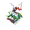
Entry Database : PDB / ID : 1gwlTitle Carbohydrate binding module family29 complexed with mannohexaose NON-CATALYTIC PROTEIN 1 Keywords / / / / Function / homology Function Domain/homology Component
/ / / / / / / / / Biological species PIROMYCES EQUI (fungus)Method / / / Resolution : 1.51 Å Authors Charnock, S.J. / Nurizzo, D. / Davies, G.J. Journal : Proc.Natl.Acad.Sci.USA / Year : 2002Title : Promiscuity in Ligand-Binding: The Three-Dimensional Structure of a Piromyces Carbohydrate-Binding Module,Cbm29-2,in Complex with Cello- and MannohexaoseAuthors : Charnock, S.J. / Bolam, D. / Nurizzo, D. / Szabo, L. / Mckie, V. / Gilbert, H. / Davies, G.J. History Deposition Mar 19, 2002 Deposition site / Processing site Revision 1.0 Mar 20, 2003 Provider / Type Revision 1.1 Jul 13, 2011 Group / Atomic model / Version format complianceRevision 2.0 Jul 29, 2020 Group Atomic model / Data collection ... Atomic model / Data collection / Derived calculations / Other / Structure summary Category atom_site / chem_comp ... atom_site / chem_comp / entity / pdbx_branch_scheme / pdbx_chem_comp_identifier / pdbx_database_status / pdbx_entity_branch / pdbx_entity_branch_descriptor / pdbx_entity_branch_link / pdbx_entity_branch_list / pdbx_entity_nonpoly / pdbx_nonpoly_scheme / pdbx_struct_assembly_gen / struct_asym / struct_conn / struct_site / struct_site_gen Item _atom_site.auth_asym_id / _atom_site.auth_seq_id ... _atom_site.auth_asym_id / _atom_site.auth_seq_id / _atom_site.label_asym_id / _chem_comp.name / _chem_comp.type / _entity.formula_weight / _entity.pdbx_description / _entity.pdbx_number_of_molecules / _entity.type / _pdbx_database_status.status_code_sf / _pdbx_struct_assembly_gen.asym_id_list / _struct_conn.pdbx_leaving_atom_flag / _struct_conn.ptnr1_auth_asym_id / _struct_conn.ptnr1_auth_seq_id / _struct_conn.ptnr1_label_asym_id / _struct_conn.ptnr2_auth_asym_id / _struct_conn.ptnr2_auth_seq_id / _struct_conn.ptnr2_label_asym_id Description / Provider / Type Revision 2.1 May 1, 2024 Group Data collection / Database references ... Data collection / Database references / Refinement description / Structure summary Category chem_comp / chem_comp_atom ... chem_comp / chem_comp_atom / chem_comp_bond / database_2 / pdbx_initial_refinement_model Item / _database_2.pdbx_DOI / _database_2.pdbx_database_accession
Show all Show less Remark 700 SHEET THE SHEET STRUCTURE OF THIS MOLECULE IS BIFURCATED. IN ORDER TO REPRESENT THIS FEATURE IN ... SHEET THE SHEET STRUCTURE OF THIS MOLECULE IS BIFURCATED. IN ORDER TO REPRESENT THIS FEATURE IN THE SHEET RECORDS BELOW, TWO SHEETS ARE DEFINED.
 Movie
Movie Controller


 Open data
 Basic information
 Components
 Keywords
 GLUCOMANNAN / CELLOHEXAOSE / MANNOHEXAOSE /
  Function and homology information
 PIROMYCES EQUI (fungus)
 Authors
 Citation
 Structure visualization
 Downloads & links
 Other downloads
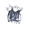


 PDBj
PDBj


 Assembly


 Mass: 18.015 Da / Num. of mol.: 200 / Source method: isolated from a natural source / Formula: H2O
 Sample preparation
 / Beamline: PX9.6 / Wavelength: 0.87
  Processing