 Movie
Movie Controller

[English] 日本語
 Yorodumi
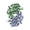
- PDB-1gph: STRUCTURE OF THE ALLOSTERIC REGULATORY ENZYME OF PURINE BIOSYNTHESIS -
+ Open data

- Basic information
| Entry | Database: PDB / ID: 1gph | ||||||
|---|---|---|---|---|---|---|---|
| Title | STRUCTURE OF THE ALLOSTERIC REGULATORY ENZYME OF PURINE BIOSYNTHESIS | ||||||
 Components Components | GLUTAMINE PHOSPHORIBOSYL-PYROPHOSPHATE AMIDOTRANSFERASE | ||||||
 Keywords Keywords |  TRANSFERASE / GLUTAMINE AMIDOTRANSFERASE TRANSFERASE / GLUTAMINE AMIDOTRANSFERASE | ||||||
| Function / homology |  Function and homology informationamidophosphoribosyltransferase / amidophosphoribosyltransferase activity / purine nucleobase biosynthetic process / purine nucleotide biosynthetic process / 'de novo' IMP biosynthetic process / glutamine metabolic process / 4 iron, 4 sulfur cluster binding / magnesium ion binding / cytoplasm Function and homology informationamidophosphoribosyltransferase / amidophosphoribosyltransferase activity / purine nucleobase biosynthetic process / purine nucleotide biosynthetic process / 'de novo' IMP biosynthetic process / glutamine metabolic process / 4 iron, 4 sulfur cluster binding / magnesium ion binding / cytoplasmSimilarity search - Function | ||||||
| Biological species |  Bacillus subtilis (bacteria) Bacillus subtilis (bacteria) | ||||||
| Method | X-RAY DIFFRACTION / Resolution: 3 Å | ||||||
 Authors Authors | Smith, J.L. | ||||||
 Citation Citation | Journal: Science / Year: 1994 Title: Structure of the allosteric regulatory enzyme of purine biosynthesis. Authors: Smith, J.L. / Zaluzec, E.J. / Wery, J.P. / Niu, L. / Switzer, R.L. / Zalkin, H. / Satow, Y. | ||||||
| History |
|
- Structure visualization
| Structure viewer | Molecule: MolmilJmol/JSmol |
|---|
- Downloads & links
-Download
| PDBx/mmCIF format | 1gph.cif.gz | 331.8 KB | Display | PDBx/mmCIF format |
|---|---|---|---|---|
| PDB format | pdb1gph.ent.gz | 274.8 KB | Display | PDB format |
| PDBx/mmJSON format | 1gph.json.gz | Tree view | PDBx/mmJSON format | |
| Others |  Other downloads Other downloads |
-Validation report
| Arichive directory | https://data.pdbj.org/pub/pdb/validation_reports/gp/1gphftp://data.pdbj.org/pub/pdb/validation_reports/gp/1gph | HTTPS FTP |
|---|
-Related structure data
| Similar structure data |
|---|


-Links
 PDBj
PDBj


- Assembly
| Deposited unit | 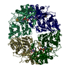
| ||||||||||||||||
|---|---|---|---|---|---|---|---|---|---|---|---|---|---|---|---|---|---|
| 1 |
| ||||||||||||||||
| 2 | 
| ||||||||||||||||
| 3 | 
| ||||||||||||||||
| Unit cell |
| ||||||||||||||||
| Atom site foot note | 1: CIS PROLINE - PRO 1 86 / 2: CIS PROLINE - PRO 2 86 / 3: CIS PROLINE - PRO 3 86 / 4: CIS PROLINE - PRO 4 86 | ||||||||||||||||
| Noncrystallographic symmetry (NCS) | NCS oper:
|
-Components
| #1: Protein | Mass: 50507.203 Da / Num. of mol.: 4 Source method: isolated from a genetically manipulated source Source: (gene. exp.) Bacillus subtilis (bacteria) / References: UniProt: P00497, amidophosphoribosyltransferase#2: Chemical | ChemComp-SF4 / Iron–sulfur cluster  Mass: 351.640 Da / Num. of mol.: 4 / Source method: obtained synthetically / Formula: Fe4S4 Mass: 351.640 Da / Num. of mol.: 4 / Source method: obtained synthetically / Formula: Fe4S4#3: Chemical | ChemComp-AMP / Adenosine monophosphate  Mass: 347.221 Da / Num. of mol.: 8 / Source method: obtained synthetically / Formula: C10H14N5O7P / Comment: AMP*YM Mass: 347.221 Da / Num. of mol.: 8 / Source method: obtained synthetically / Formula: C10H14N5O7P / Comment: AMP*YMNonpolymer details | THE FE ATOMS OF THE [4FE-4S] CLUSTER ARE LIGATED BY CYSTEINE S(GAMMA) ATOMS AS FOLLOWS: FE 1 - SG ...THE FE ATOMS OF THE [4FE-4S] CLUSTER ARE LIGATED BY CYSTEINE S(GAMMA) ATOMS AS FOLLOWS: FE 1 - SG CYS 437; FE 2 - SG CYS 236; FE 3 - SG CYS 440; FE 4 - SG CYS 382. THE AMP MOLECULE THAT IS RESIDUE 467 IS BOUND TO THE SITE WHERE THE SUBSTRATE PRPP IS PRESUMED TO BIND. THE AMP MOLECULE THAT IS RESIDUE 468 IS BOUND BETWEEN SUBUNITS IN WHAT IS PRESUMED TO BE AN ALLOSTERIC | Sequence details | THE STRUCTURE DIFFERS FROM THE DEPOSITED SEQUENCE IN TWO PLACES: 1) AN 11-RESIDUE PROPEPTIDE ...THE STRUCTURE DIFFERS FROM THE DEPOSITED SEQUENCE IN TWO PLACES: 1) AN 11-RESIDUE PROPEPTIDE | |
|---|
-Experimental details
-Experiment
| Experiment | Method: X-RAY DIFFRACTION |
|---|
- Sample preparation
| Crystal | Density Matthews: 2.8 Å3/Da / Density % sol: 56.04 % | ||||||||||||||||||||||||||||||||||||||||||||||||||||||||||||||||||||||||
|---|---|---|---|---|---|---|---|---|---|---|---|---|---|---|---|---|---|---|---|---|---|---|---|---|---|---|---|---|---|---|---|---|---|---|---|---|---|---|---|---|---|---|---|---|---|---|---|---|---|---|---|---|---|---|---|---|---|---|---|---|---|---|---|---|---|---|---|---|---|---|---|---|---|
| Crystal grow | *PLUS pH: 7.9 / Method: batch method | ||||||||||||||||||||||||||||||||||||||||||||||||||||||||||||||||||||||||
| Components of the solutions | *PLUS
|
-Data collection
| Radiation | Scattering type: x-ray |
|---|---|
| Radiation wavelength | Relative weight: 1 |
| Reflection | *PLUS Highest resolution: 3 Å / Num. obs: 41267 / % possible obs: 90.1 % |
- Processing
| Software |
| ||||||||||||||||||||||||||||||||||||||||||||||||||||||||||||
|---|---|---|---|---|---|---|---|---|---|---|---|---|---|---|---|---|---|---|---|---|---|---|---|---|---|---|---|---|---|---|---|---|---|---|---|---|---|---|---|---|---|---|---|---|---|---|---|---|---|---|---|---|---|---|---|---|---|---|---|---|---|
| Refinement | Resolution: 3→7 Å / σ(F): 2 /
| ||||||||||||||||||||||||||||||||||||||||||||||||||||||||||||
| Refinement step | Cycle: LAST / Resolution: 3→7 Å
| ||||||||||||||||||||||||||||||||||||||||||||||||||||||||||||
| Refine LS restraints |
| ||||||||||||||||||||||||||||||||||||||||||||||||||||||||||||
| Software | *PLUS Name: X-PLOR / Classification: refinement | ||||||||||||||||||||||||||||||||||||||||||||||||||||||||||||
| Refinement | *PLUS Rfactor obs: 0.182 / Rfactor Rwork: 0.182 | ||||||||||||||||||||||||||||||||||||||||||||||||||||||||||||
| Solvent computation | *PLUS | ||||||||||||||||||||||||||||||||||||||||||||||||||||||||||||
| Displacement parameters | *PLUS | ||||||||||||||||||||||||||||||||||||||||||||||||||||||||||||
| Refine LS restraints | *PLUS Type: x_angle_d / Dev ideal: 3.3 |
