 Movie
Movie Controller


+ Open data

- Basic information
| Entry | Database: PDB / ID: 1eh7 | ||||||
|---|---|---|---|---|---|---|---|
| Title | METHYLATED HUMAN O6-ALKYLGUANINE-DNA ALKYLTRANSFERASE | ||||||
 Components Components | O6-ALKYLGUANINE-DNA ALKYLTRANSFERASE | ||||||
 Keywords Keywords |  TRANSFERASE / ALKYLTRANSFERASE / METHYLTRANSFERASE / DNA REPAIR TRANSFERASE / ALKYLTRANSFERASE / METHYLTRANSFERASE / DNA REPAIR | ||||||
| Function / homology |  Function and homology information Function and homology informationMGMT-mediated DNA damage reversal / methylated-DNA-[protein]-cysteine S-methyltransferase / methylated-DNA-[protein]-cysteine S-methyltransferase activity / DNA-methyltransferase activity / DNA alkylation repair / DNA ligation / positive regulation of double-strand break repair / methyltransferase activity / DNA repair / negative regulation of apoptotic process ...MGMT-mediated DNA damage reversal / methylated-DNA-[protein]-cysteine S-methyltransferase / methylated-DNA-[protein]-cysteine S-methyltransferase activity / DNA-methyltransferase activity / DNA alkylation repair / DNA ligation / positive regulation of double-strand break repair / methyltransferase activity / DNA repair / negative regulation of apoptotic process / DNA binding / nucleoplasm / membrane / metal ion binding / nucleusSimilarity search - Function | ||||||
| Biological species |  Homo sapiens (human) Homo sapiens (human) | ||||||
| Method | X-RAY DIFFRACTION / SYNCHROTRON / Resolution: 2 Å | ||||||
 Authors Authors | Daniels, D.S. / Tainer, J.A. | ||||||
 Citation Citation | Journal: EMBO J. / Year: 2000 Title: Active and alkylated human AGT structures: a novel zinc site, inhibitor and extrahelical base binding. Authors: Daniels, D.S. / Mol, C.D. / Arvai, A.S. / Kanugula, S. / Pegg, A.E. / Tainer, J.A. | ||||||
| History |
|
- Structure visualization
| Structure viewer | Molecule: MolmilJmol/JSmol |
|---|
- Downloads & links
-Download
| PDBx/mmCIF format | 1eh7.cif.gz | 49.8 KB | Display | PDBx/mmCIF format |
|---|---|---|---|---|
| PDB format | pdb1eh7.ent.gz | 33.4 KB | Display | PDB format |
| PDBx/mmJSON format | 1eh7.json.gz | Tree view | PDBx/mmJSON format | |
| Others |  Other downloads Other downloads |
-Validation report
| Arichive directory | https://data.pdbj.org/pub/pdb/validation_reports/eh/1eh7ftp://data.pdbj.org/pub/pdb/validation_reports/eh/1eh7 | HTTPS FTP |
|---|
-Related structure data
| Related structure data | 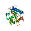 1eh6SC  1eh8C S: Starting model for refinement C: citing same article ( |
|---|---|
| Similar structure data |


-Links
 PDBj
PDBj- Assembly
| Deposited unit | 
| ||||||||
|---|---|---|---|---|---|---|---|---|---|
| 1 |
| ||||||||
| Unit cell |
|
-Components
| #1: Protein | Mass: 21685.977 Da / Num. of mol.: 1 Source method: isolated from a genetically manipulated source Details: METHYLATED CYSTEINE 145 / Source: (gene. exp.) Homo sapiens (human) / Production host:  Escherichia coli (E. coli) Escherichia coli (E. coli)References: UniProt: P16455, methylated-DNA-[protein]-cysteine S-methyltransferase |
|---|---|
| #2: Chemical | ChemComp-ZN /   Mass: 65.409 Da / Num. of mol.: 1 / Source method: obtained synthetically / Formula: Zn Mass: 65.409 Da / Num. of mol.: 1 / Source method: obtained synthetically / Formula: Zn |
| #3: Water | ChemComp-HOH / Water Mass: 18.015 Da / Num. of mol.: 182 / Source method: isolated from a natural source / Formula: H2O Mass: 18.015 Da / Num. of mol.: 182 / Source method: isolated from a natural source / Formula: H2O |
-Experimental details
-Experiment
| Experiment | Method: X-RAY DIFFRACTION / Number of used crystals: 1 |
|---|
- Sample preparation
| Crystal | Density Matthews: 2.59 Å3/Da / Density % sol: 52.47 % | |||||||||||||||
|---|---|---|---|---|---|---|---|---|---|---|---|---|---|---|---|---|
| Crystal grow | Temperature: 298 K / Method: vapor diffusion, hanging drop / pH: 7.5 Details: 18 MG/ML PROTEIN, 1.5 M SUCROSE, pH 7.5, VAPOR DIFFUSION, HANGING DROP, temperature 298K | |||||||||||||||
| Crystal grow | *PLUS Temperature: 25 ℃ | |||||||||||||||
| Components of the solutions | *PLUS
|
-Data collection
| Diffraction | Mean temperature: 100 K |
|---|---|
| Diffraction source | Source: SYNCHROTRON / Site: SSRL  / Beamline: BL7-1 / Wavelength: 1.08 / Beamline: BL7-1 / Wavelength: 1.08 |
| Detector | Type: MARRESEARCH / Detector: IMAGE PLATE / Date: Mar 16, 1999 |
| Radiation | Protocol: SINGLE WAVELENGTH / Monochromatic (M) / Laue (L): M / Scattering type: x-ray |
| Radiation wavelength | Wavelength: 1.08 Å / Relative weight: 1 |
| Reflection | Resolution: 2→20 Å / Num. all: 28548 / Num. obs: 15195 / % possible obs: 97.2 % / Observed criterion σ(I): -3 / Redundancy: 1.472 % / Biso Wilson estimate: 14.9 Å2 / Rmerge(I) obs: 0.051 / Net I/σ(I): 13.4926 |
| Reflection shell | Resolution: 2→2.07 Å / Redundancy: 1.858 % / Rmerge(I) obs: 0.255 / Mean I/σ(I) obs: 3.133 / % possible all: 98.4 |
| Reflection | *PLUS Highest resolution: 2 Å / Num. measured all: 28548 |
| Reflection shell | *PLUS % possible obs: 98.2 % |
- Processing
| Software |
| ||||||||||||||||||||||||||||||||||||||||||||||||||||||||||||||||||||||||||||||||
|---|---|---|---|---|---|---|---|---|---|---|---|---|---|---|---|---|---|---|---|---|---|---|---|---|---|---|---|---|---|---|---|---|---|---|---|---|---|---|---|---|---|---|---|---|---|---|---|---|---|---|---|---|---|---|---|---|---|---|---|---|---|---|---|---|---|---|---|---|---|---|---|---|---|---|---|---|---|---|---|---|---|
| Refinement | Starting model: PDB 1EH6 Resolution: 2→18.23 Å / Rfactor Rfree error: 0.006 / Data cutoff high absF: 1710146.25 / Data cutoff low absF: 0 / Isotropic thermal model: RESTRAINED / Cross valid method: THROUGHOUT / σ(F): 0 / Stereochemistry target values: Engh & Huber Details: ILE 141 IS LOCATED IN A TIGHT TURN PRECEDING THE ACTIVE SITE AND CONSTRAINED IN A DISALLOWED RAMACHANDRAN REGION.
| ||||||||||||||||||||||||||||||||||||||||||||||||||||||||||||||||||||||||||||||||
| Solvent computation | Solvent model: FLAT MODEL / Bsol: 76.82 Å2 / ksol: 0.473 e/Å3 | ||||||||||||||||||||||||||||||||||||||||||||||||||||||||||||||||||||||||||||||||
| Displacement parameters | Biso mean: 26.9 Å2
| ||||||||||||||||||||||||||||||||||||||||||||||||||||||||||||||||||||||||||||||||
| Refine analyze |
| ||||||||||||||||||||||||||||||||||||||||||||||||||||||||||||||||||||||||||||||||
| Refinement step | Cycle: LAST / Resolution: 2→18.23 Å
| ||||||||||||||||||||||||||||||||||||||||||||||||||||||||||||||||||||||||||||||||
| Refine LS restraints |
| ||||||||||||||||||||||||||||||||||||||||||||||||||||||||||||||||||||||||||||||||
| LS refinement shell | Resolution: 2→2.13 Å / Rfactor Rfree error: 0.018 / Total num. of bins used: 6
| ||||||||||||||||||||||||||||||||||||||||||||||||||||||||||||||||||||||||||||||||
| Xplor file |
| ||||||||||||||||||||||||||||||||||||||||||||||||||||||||||||||||||||||||||||||||
| Software | *PLUS Name: CNS / Version: 0.9 / Classification: refinement | ||||||||||||||||||||||||||||||||||||||||||||||||||||||||||||||||||||||||||||||||
| Refinement | *PLUS σ(F): 0 / % reflection Rfree: 10.1 % / Rfactor obs: 0.199 | ||||||||||||||||||||||||||||||||||||||||||||||||||||||||||||||||||||||||||||||||
| Solvent computation | *PLUS | ||||||||||||||||||||||||||||||||||||||||||||||||||||||||||||||||||||||||||||||||
| Displacement parameters | *PLUS Biso mean: 26.9 Å2 | ||||||||||||||||||||||||||||||||||||||||||||||||||||||||||||||||||||||||||||||||
| Refine LS restraints | *PLUS
| ||||||||||||||||||||||||||||||||||||||||||||||||||||||||||||||||||||||||||||||||
| LS refinement shell | *PLUS Rfactor Rfree: 0.284 / % reflection Rfree: 9.8 % |
