 Movie
Movie Controller

[English] 日本語
 Yorodumi
- PDB-1e92: Pteridine reductase 1 from Leishmania major complexed with NADP+ ... -
+ Open data

- Basic information
| Entry | Database: PDB / ID: 1.0E+92 | ||||||
|---|---|---|---|---|---|---|---|
| Title | Pteridine reductase 1 from Leishmania major complexed with NADP+ and dihydrobiopterin | ||||||
 Components Components | PTERIDINE REDUCTASE 1 | ||||||
 Keywords Keywords | PTERIDINE REDUCTASE / TRYPANOSOMATIDS / DRUG RESISTANCE / PTERIN SALVAGE / SHORT-CHAIN DEHYDROGENASE/REDUCTASE | ||||||
| Function / homology |  Function and homology informationpteridine reductase / 6,7-dihydropteridine reductase activity / pteridine reductase activity / tetrahydrobiopterin biosynthetic process / response to methotrexate / oxidoreductase activity / cytosol Function and homology informationpteridine reductase / 6,7-dihydropteridine reductase activity / pteridine reductase activity / tetrahydrobiopterin biosynthetic process / response to methotrexate / oxidoreductase activity / cytosolSimilarity search - Function | ||||||
| Biological species |  LEISHMANIA MAJOR (eukaryote) LEISHMANIA MAJOR (eukaryote) | ||||||
| Method | X-RAY DIFFRACTION / SYNCHROTRON / MOLECULAR REPLACEMENT / Resolution: 2.2 Å | ||||||
 Authors Authors | Schuettelkopf, A.W. / Hunter, W.N. | ||||||
 Citation Citation | Journal: Nat.Struct.Biol. / Year: 2001 Title: Pteridine Reductase Mechanism Correlates Pterin Metabolism with Drug Resistance in Trypanosomatid Parasites Authors: Gourley, D.G. / Schuettelkopf, A.W. / Leonard, G.A. / Luba, J. / Hardy, L.W. / Beverley, S.M. / Hunter, W.N. | ||||||
| History |
|
- Structure visualization
| Structure viewer | Molecule: MolmilJmol/JSmol |
|---|
- Downloads & links
-Download
| PDBx/mmCIF format | 1e92.cif.gz | 218.4 KB | Display | PDBx/mmCIF format |
|---|---|---|---|---|
| PDB format | pdb1e92.ent.gz | 175.9 KB | Display | PDB format |
| PDBx/mmJSON format | 1e92.json.gz | Tree view | PDBx/mmJSON format | |
| Others |  Other downloads Other downloads |
-Validation report
| Arichive directory | https://data.pdbj.org/pub/pdb/validation_reports/e9/1e92ftp://data.pdbj.org/pub/pdb/validation_reports/e9/1e92 | HTTPS FTP |
|---|
-Related structure data
| Related structure data | 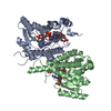 1e7wSC S: Starting model for refinement C: citing same article ( |
|---|---|
| Similar structure data |


-Links
 PDBj
PDBj


- Assembly
| Deposited unit | 
| ||||||||||||||||
|---|---|---|---|---|---|---|---|---|---|---|---|---|---|---|---|---|---|
| 1 |
| ||||||||||||||||
| Unit cell |
| ||||||||||||||||
| Noncrystallographic symmetry (NCS) | NCS oper:
|
-Components
| #1: Protein | / PTR1 Mass: 30440.580 Da / Num. of mol.: 4 Source method: isolated from a genetically manipulated source Source: (gene. exp.) LEISHMANIA MAJOR (eukaryote) / Gene: PTR1 / Plasmid: PET 15B / Production host:  ESCHERICHIA COLI (E. coli) / Strain (production host): BL21(DE3) ESCHERICHIA COLI (E. coli) / Strain (production host): BL21(DE3)References: TrEMBL: Q9U1F8, UniProt: Q01782*PLUS, EC: 1.1.1.253#2: Chemical | ChemComp-NAP / Nicotinamide adenine dinucleotide phosphate  Mass: 743.405 Da / Num. of mol.: 4 / Source method: obtained synthetically / Formula: C21H28N7O17P3 Mass: 743.405 Da / Num. of mol.: 4 / Source method: obtained synthetically / Formula: C21H28N7O17P3#3: Chemical | ChemComp-HBI / Dihydrobiopterin  Mass: 239.231 Da / Num. of mol.: 4 / Source method: obtained synthetically / Formula: C9H13N5O3 Mass: 239.231 Da / Num. of mol.: 4 / Source method: obtained synthetically / Formula: C9H13N5O3#4: Chemical | ChemComp-EDO / Ethylene glycol  Mass: 62.068 Da / Num. of mol.: 4 / Source method: obtained synthetically / Formula: C2H6O2 Mass: 62.068 Da / Num. of mol.: 4 / Source method: obtained synthetically / Formula: C2H6O2#5: Water | ChemComp-HOH / | Water Mass: 18.015 Da / Num. of mol.: 434 / Source method: isolated from a natural source / Formula: H2O Mass: 18.015 Da / Num. of mol.: 434 / Source method: isolated from a natural source / Formula: H2O |
|---|
-Experimental details
-Experiment
| Experiment | Method: X-RAY DIFFRACTION / Number of used crystals: 1 |
|---|
- Sample preparation
| Crystal | Density Matthews: 2.8 Å3/Da / Density % sol: 56 % | |||||||||||||||||||||||||||||||||||||||||||||
|---|---|---|---|---|---|---|---|---|---|---|---|---|---|---|---|---|---|---|---|---|---|---|---|---|---|---|---|---|---|---|---|---|---|---|---|---|---|---|---|---|---|---|---|---|---|---|
| Crystal grow | Temperature: 293 K / pH: 5.4 Details: 15 MG/ML PROTEIN, 0.1M NAAC/HAC BUFFER PH 5.5, 12% MPEG 5000, 0.075M CAAC2, 293K | |||||||||||||||||||||||||||||||||||||||||||||
| Crystal grow | *PLUS Temperature: 293 K / pH: 7 / Method: vapor diffusion, sitting dropDetails: Gourley, D.G., (1999) Acta Crystallogr., D55, 1608. | |||||||||||||||||||||||||||||||||||||||||||||
| Components of the solutions | *PLUS
|
-Data collection
| Diffraction | Mean temperature: 100 K |
|---|---|
| Diffraction source | Source: SYNCHROTRON / Site: ESRF  / Beamline: ID14-2 / Wavelength: 0.9326 / Beamline: ID14-2 / Wavelength: 0.9326 |
| Detector | Type: MARRESEARCH / Detector: CCD |
| Radiation | Protocol: SINGLE WAVELENGTH / Monochromatic (M) / Laue (L): M / Scattering type: x-ray |
| Radiation wavelength | Wavelength: 0.9326 Å / Relative weight: 1 |
| Reflection | Resolution: 2.2→27.74 Å / Num. obs: 62471 / % possible obs: 91.1 % / Redundancy: 2.4 % / Biso Wilson estimate: 25.3 Å2 / Rsym value: 0.077 / Net I/σ(I): 18.9 |
| Reflection shell | Resolution: 2.2→2.25 Å / Redundancy: 2.2 % / Mean I/σ(I) obs: 6.7 / Rsym value: 0.198 / % possible all: 84.5 |
| Reflection | *PLUS Num. measured all: 449654 / Rmerge(I) obs: 0.077 |
| Reflection shell | *PLUS Rmerge(I) obs: 0.198 |
- Processing
| Software |
| ||||||||||||||||||||||||||||||||||||||||||||||||||||||||||||||||||||||||||||||||
|---|---|---|---|---|---|---|---|---|---|---|---|---|---|---|---|---|---|---|---|---|---|---|---|---|---|---|---|---|---|---|---|---|---|---|---|---|---|---|---|---|---|---|---|---|---|---|---|---|---|---|---|---|---|---|---|---|---|---|---|---|---|---|---|---|---|---|---|---|---|---|---|---|---|---|---|---|---|---|---|---|---|
| Refinement | Method to determine structure: MOLECULAR REPLACEMENT Starting model: PDB ENTRY 1E7W Resolution: 2.2→27.77 Å / Rfactor Rfree error: 0.004 / Data cutoff high absF: 2824839.6 / Isotropic thermal model: RESTRAINED / Cross valid method: THROUGHOUT / σ(F): 0 Details: THE SIDE CHAIN OF ARG A20 WAS MODELLED WITH TWO ALTERNATE CONFORMATIONS, EACH OF HALF OCCUPANCY. FOR THE SIDE CHAIN ATOMS (OTHER THAN CB) OF RESIDUES A122, A133, A136, A168, C120, C121, ...Details: THE SIDE CHAIN OF ARG A20 WAS MODELLED WITH TWO ALTERNATE CONFORMATIONS, EACH OF HALF OCCUPANCY. FOR THE SIDE CHAIN ATOMS (OTHER THAN CB) OF RESIDUES A122, A133, A136, A168, C120, C121, C132, C133, C137, D120, D168 THE OCCUPANCY VALUES WERE SET TO 0.00 DUE TO LACK OF INTERPRETABLE DENSITY.
| ||||||||||||||||||||||||||||||||||||||||||||||||||||||||||||||||||||||||||||||||
| Solvent computation | Solvent model: FLAT MODEL / Bsol: 30.0631 Å2 / ksol: 0.341437 e/Å3 | ||||||||||||||||||||||||||||||||||||||||||||||||||||||||||||||||||||||||||||||||
| Displacement parameters | Biso mean: 23.7 Å2
| ||||||||||||||||||||||||||||||||||||||||||||||||||||||||||||||||||||||||||||||||
| Refine analyze |
| ||||||||||||||||||||||||||||||||||||||||||||||||||||||||||||||||||||||||||||||||
| Refinement step | Cycle: LAST / Resolution: 2.2→27.77 Å
| ||||||||||||||||||||||||||||||||||||||||||||||||||||||||||||||||||||||||||||||||
| Refine LS restraints |
| ||||||||||||||||||||||||||||||||||||||||||||||||||||||||||||||||||||||||||||||||
| LS refinement shell | Resolution: 2.2→2.34 Å / Rfactor Rfree error: 0.011 / Total num. of bins used: 6
| ||||||||||||||||||||||||||||||||||||||||||||||||||||||||||||||||||||||||||||||||
| Xplor file |
| ||||||||||||||||||||||||||||||||||||||||||||||||||||||||||||||||||||||||||||||||
| Software | *PLUS Name: CNS / Version: 1 / Classification: refinement | ||||||||||||||||||||||||||||||||||||||||||||||||||||||||||||||||||||||||||||||||
| Refine LS restraints | *PLUS
| ||||||||||||||||||||||||||||||||||||||||||||||||||||||||||||||||||||||||||||||||
| LS refinement shell | *PLUS Rfactor Rwork: 0.2 |
