 Movie
Movie Controller

[English] 日本語
 Yorodumi
- PDB-1dgk: MUTANT MONOMER OF RECOMBINANT HUMAN HEXOKINASE TYPE I WITH GLUCOS... -
+ Open data

- Basic information
| Entry | Database: PDB / ID: 1dgk | ||||||
|---|---|---|---|---|---|---|---|
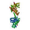
| Title | MUTANT MONOMER OF RECOMBINANT HUMAN HEXOKINASE TYPE I WITH GLUCOSE AND ADP IN THE ACTIVE SITE | ||||||
 Components Components | HEXOKINASE TYPE I | ||||||
 Keywords Keywords |  TRANSFERASE / BRAIN HEXOKINASE / MAMMALIAN HEXOKINASE 1 / SUGAR KINASE TRANSFERASE / BRAIN HEXOKINASE / MAMMALIAN HEXOKINASE 1 / SUGAR KINASE | ||||||
| Function / homology |  Function and homology information Function and homology informationDefective HK1 causes hexokinase deficiency (HK deficiency) / glucosamine kinase activity / hexokinase activity / mannokinase activity / maintenance of protein location in mitochondrion / positive regulation of cytokine production involved in immune response / establishment of protein localization to mitochondrion / hexokinase / fructokinase activity / carbohydrate phosphorylation ...Defective HK1 causes hexokinase deficiency (HK deficiency) / glucosamine kinase activity / hexokinase activity / mannokinase activity / maintenance of protein location in mitochondrion / positive regulation of cytokine production involved in immune response / establishment of protein localization to mitochondrion / hexokinase / fructokinase activity / carbohydrate phosphorylation / glucokinase activity / mannose metabolic process / glucose 6-phosphate metabolic process / peptidoglycan binding / D-glucose binding / fructose 6-phosphate metabolic process / canonical glycolysis / Glycolysis / intracellular glucose homeostasis / positive regulation of interleukin-1 beta production / glycolytic process / glucose metabolic process / mitochondrial outer membrane / inflammatory response / membrane raft / innate immune response / mitochondrion / ATP binding / cytosolSimilarity search - Function | ||||||
| Biological species |  Homo sapiens (human) Homo sapiens (human) | ||||||
| Method | X-RAY DIFFRACTION / SYNCHROTRON / Resolution: 2.8 Å | ||||||
 Authors Authors | Aleshin, A.E. / Liu, X. / Kirby, C. / Bourenkov, G.P. / Bartunik, H.D. / Fromm, H.J. / Honzatko, R.B. | ||||||
 Citation Citation | Journal: J.Mol.Biol. / Year: 2000 Title: Crystal structures of mutant monomeric hexokinase I reveal multiple ADP binding sites and conformational changes relevant to allosteric regulation. Authors: Aleshin, A.E. / Kirby, C. / Liu, X. / Bourenkov, G.P. / Bartunik, H.D. / Fromm, H.J. / Honzatko, R.B. #1: Journal: J.Mol.Biol. / Year: 1998Title: Regulation of Hexokinase I: Crystal Structure of Recombinant Human Brain Hexokinase Complexed with Glucose and Phosphate Authors: Aleshin, A.E. / Zeng, C. / Bartunik, H.D. / Fromm, H.J. / Honzatko, R.B. #2: Journal: Structure / Year: 1998Title: The Mechanism of Regulation of Hexokinase: New Insights from the Crystal Structure of Recombinant Human Brain Hexokinase Complexed with Glucose and Glucose-6-Phosphate Authors: Aleshin, A.E. / Zeng, C. / Bartunik, H.D. / Fromm, H.J. / Honzatko, R.B. | ||||||
| History |
|
- Structure visualization
| Structure viewer | Molecule: MolmilJmol/JSmol |
|---|
- Downloads & links
-Download
| PDBx/mmCIF format | 1dgk.cif.gz | 192 KB | Display | PDBx/mmCIF format |
|---|---|---|---|---|
| PDB format | pdb1dgk.ent.gz | 151 KB | Display | PDB format |
| PDBx/mmJSON format | 1dgk.json.gz | Tree view | PDBx/mmJSON format | |
| Others |  Other downloads Other downloads |
-Validation report
| Arichive directory | https://data.pdbj.org/pub/pdb/validation_reports/dg/1dgkftp://data.pdbj.org/pub/pdb/validation_reports/dg/1dgk | HTTPS FTP |
|---|
-Related structure data


-Links
 PDBj
PDBj


- Assembly
| Deposited unit | 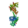
| ||||||||
|---|---|---|---|---|---|---|---|---|---|
| 1 |
| ||||||||
| Unit cell |
|
-Components
| #1: Protein | Mass: 102556.984 Da / Num. of mol.: 1 / Mutation: E280A, R283A, G284Y, T536A Source method: isolated from a genetically manipulated source Details: SECOND MOLECULE OF ADP IS BOUND NEAR THE N-TERMINUS OF THE POLYPEPTIDE CHAIN. THE REGULATORY SITE OF THE N-TERMINAL DOMAIN IS OCCUPIED WITH GLUCOSE AND PHOSPHATE. MUTATIONS IN DIMER INTERFACE AND THE ACTIVE SITE Source: (gene. exp.) Homo sapiens (human) / Cell: NEURON / Organ: BRAIN / Plasmid: PET11A / Production host:  Escherichia coli (E. coli) / References: UniProt: P19367, hexokinase Escherichia coli (E. coli) / References: UniProt: P19367, hexokinase | ||||||
|---|---|---|---|---|---|---|---|
| #2: Sugar | Glucose  Type: D-saccharide, alpha linking / Mass: 180.156 Da / Num. of mol.: 2 / Source method: obtained synthetically / Formula: C6H12O6 Type: D-saccharide, alpha linking / Mass: 180.156 Da / Num. of mol.: 2 / Source method: obtained synthetically / Formula: C6H12O6#3: Chemical | ChemComp-PO4 / | Phosphate  Mass: 94.971 Da / Num. of mol.: 1 / Source method: obtained synthetically / Formula: PO4 Mass: 94.971 Da / Num. of mol.: 1 / Source method: obtained synthetically / Formula: PO4#4: Chemical | Adenosine diphosphate  Mass: 427.201 Da / Num. of mol.: 2 / Source method: obtained synthetically / Formula: C10H15N5O10P2 / Comment: ADP, energy-carrying molecule*YM Mass: 427.201 Da / Num. of mol.: 2 / Source method: obtained synthetically / Formula: C10H15N5O10P2 / Comment: ADP, energy-carrying molecule*YM#5: Water | ChemComp-HOH / | Water Mass: 18.015 Da / Num. of mol.: 94 / Source method: isolated from a natural source / Formula: H2O Mass: 18.015 Da / Num. of mol.: 94 / Source method: isolated from a natural source / Formula: H2O |
-Experimental details
-Experiment
| Experiment | Method: X-RAY DIFFRACTION / Number of used crystals: 1 |
|---|
- Sample preparation
| Crystal | Density Matthews: 3.06 Å3/Da / Density % sol: 59.76 % | |||||||||||||||||||||||||||||||||||||||||||||||||||||||||||||||
|---|---|---|---|---|---|---|---|---|---|---|---|---|---|---|---|---|---|---|---|---|---|---|---|---|---|---|---|---|---|---|---|---|---|---|---|---|---|---|---|---|---|---|---|---|---|---|---|---|---|---|---|---|---|---|---|---|---|---|---|---|---|---|---|---|
| Crystal grow | Temperature: 298 K / Method: vapor diffusion, hanging drop / pH: 6.5 Details: PEG 8000, SODIUM ACETATE, MES, ADP, GLUCOSE, GLUCOSE-6-PHOSPHATE. GROWN CRYSTALS WERE SOAKED WITH THE SAME BUFFER, BUT W/O GLUCOSE-6-PHOSPHATE, pH 6.5, VAPOR DIFFUSION, HANGING DROP, temperature 298K | |||||||||||||||||||||||||||||||||||||||||||||||||||||||||||||||
| Crystal grow | *PLUS Details: drop consists of equal volume of protein and precipitant solutionsPH range low: 6.5 / PH range high: 5.8 | |||||||||||||||||||||||||||||||||||||||||||||||||||||||||||||||
| Components of the solutions | *PLUS
|
-Data collection
| Diffraction | Mean temperature: 110 K |
|---|---|
| Diffraction source | Source: SYNCHROTRON / Site: APS  / Beamline: 14-BM-D / Wavelength: 1 / Beamline: 14-BM-D / Wavelength: 1 |
| Detector | Type: ADSC QUANTUM 4 / Detector: CCD / Date: Feb 3, 1999 |
| Radiation | Protocol: SINGLE WAVELENGTH / Monochromatic (M) / Laue (L): M / Scattering type: x-ray |
| Radiation wavelength | Wavelength: 1 Å / Relative weight: 1 |
| Reflection | Resolution: 2.77→50 Å / Num. all: 65575 / Num. obs: 25465 / % possible obs: 78 % / Redundancy: 2.64 % / Biso Wilson estimate: 54 Å2 / Rmerge(I) obs: 0.064 / Net I/σ(I): 15 |
| Reflection shell | Resolution: 2.77→2.98 Å / Redundancy: 2 % / Rmerge(I) obs: 0.46 / % possible all: 57 |
| Reflection | *PLUS Num. obs: 25491 / % possible obs: 77.5 % / Num. measured all: 65575 |
| Reflection shell | *PLUS % possible obs: 57 % / Rmerge(I) obs: 0.44 |
- Processing
| Software |
| |||||||||||||||||||||||||||||||||||||||||||||||||||||||||||||||
|---|---|---|---|---|---|---|---|---|---|---|---|---|---|---|---|---|---|---|---|---|---|---|---|---|---|---|---|---|---|---|---|---|---|---|---|---|---|---|---|---|---|---|---|---|---|---|---|---|---|---|---|---|---|---|---|---|---|---|---|---|---|---|---|---|
| Refinement | Resolution: 2.8→8 Å / σ(F): 0 / σ(I): 0 / Stereochemistry target values: ENGH & HUBER Details: USED MAXIMUM LIKELIHOOD RESIDUALS AND CONJUGATE DIRECTION MINIMIZATION.SIDE CHAINS OF RESIDUES 16, 17, 20, 21, 24, 101, 102, 252, 353, 794-799, 801, 804, 806, 809-811 HAVE POOR ELECTRON ...Details: USED MAXIMUM LIKELIHOOD RESIDUALS AND CONJUGATE DIRECTION MINIMIZATION.SIDE CHAINS OF RESIDUES 16, 17, 20, 21, 24, 101, 102, 252, 353, 794-799, 801, 804, 806, 809-811 HAVE POOR ELECTRON DENSITY. THEIR OCCUPANCIES ARE SET TO 0.01
| |||||||||||||||||||||||||||||||||||||||||||||||||||||||||||||||
| Refinement step | Cycle: LAST / Resolution: 2.8→8 Å
| |||||||||||||||||||||||||||||||||||||||||||||||||||||||||||||||
| Refine LS restraints |
| |||||||||||||||||||||||||||||||||||||||||||||||||||||||||||||||
| Software | *PLUS Name: REFMAC / Classification: refinement | |||||||||||||||||||||||||||||||||||||||||||||||||||||||||||||||
| Refinement | *PLUS Rfactor Rfree: 0.31 / Rfactor Rwork: 0.26 | |||||||||||||||||||||||||||||||||||||||||||||||||||||||||||||||
| Solvent computation | *PLUS | |||||||||||||||||||||||||||||||||||||||||||||||||||||||||||||||
| Displacement parameters | *PLUS | |||||||||||||||||||||||||||||||||||||||||||||||||||||||||||||||
| Refine LS restraints | *PLUS
|
