 Movie
Movie Controller


+ Open data

- Basic information
| Entry | Database: PDB / ID: 1a7h | ||||||
|---|---|---|---|---|---|---|---|
| Title | GAMMA S CRYSTALLIN C-TERMINAL DOMAIN | ||||||
 Components Components | GAMMAS CRYSTALLIN | ||||||
 Keywords Keywords | EYE-LENS PROTEIN / GAMMA CRYSTALLIN S /  EYE LENS PROTEIN / MULTIGENE FAMILY EYE LENS PROTEIN / MULTIGENE FAMILY | ||||||
| Function / homology |  Function and homology information Function and homology informationstructural constituent of eye lens / lens development in camera-type eye / visual perception / morphogenesis of an epitheliumSimilarity search - Function | ||||||
| Biological species |  Bos taurus (cattle) Bos taurus (cattle) | ||||||
| Method | X-RAY DIFFRACTION / SYNCHROTRON / MOLECULAR REPLACEMENT / Resolution: 2.56 Å | ||||||
 Authors Authors | Basak, A.K. / Slingsby, C. | ||||||
 Citation Citation | Journal: Protein Eng. / Year: 1998 Title: The C-terminal domains of gammaS-crystallin pair about a distorted twofold axis. Authors: Basak, A.K. / Kroone, R.C. / Lubsen, N.H. / Naylor, C.E. / Jaenicke, R. / Slingsby, C. | ||||||
| History |
|
- Structure visualization
| Structure viewer | Molecule: MolmilJmol/JSmol |
|---|
- Downloads & links
-Download
| PDBx/mmCIF format | 1a7h.cif.gz | 48.8 KB | Display | PDBx/mmCIF format |
|---|---|---|---|---|
| PDB format | pdb1a7h.ent.gz | 35.4 KB | Display | PDB format |
| PDBx/mmJSON format | 1a7h.json.gz | Tree view | PDBx/mmJSON format | |
| Others |  Other downloads Other downloads |
-Validation report
| Arichive directory | https://data.pdbj.org/pub/pdb/validation_reports/a7/1a7hftp://data.pdbj.org/pub/pdb/validation_reports/a7/1a7h | HTTPS FTP |
|---|
-Related structure data
| Similar structure data |
|---|


-Links
 PDBj
PDBj
- Assembly
| Deposited unit | 
| ||||||||
|---|---|---|---|---|---|---|---|---|---|
| 1 | 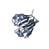
| ||||||||
| 2 | 
| ||||||||
| Unit cell |
| ||||||||
| Noncrystallographic symmetry (NCS) | NCS oper: (Code: given Matrix: (-0.35584, -0.08497, -0.93068), Vector : Details | THE PROTEIN IN THE CRYSTAL LATTICE IS A DIMER. IN DILUTE SOLUTION IT IS A MONOMER. THERE IS NO NATURALLY OCCURRING BIOLOGICAL EQUIVALENT. | |
-Components
| #1: Protein | Mass: 10319.770 Da / Num. of mol.: 2 / Fragment: C-TERMINAL DOMAIN / Mutation: Q87M Source method: isolated from a genetically manipulated source Source: (gene. exp.) Bos taurus (cattle) / Cell line: BL21 / Gene: CRYGS / Organ: EYE / Organelle: EYE-LENS / Plasmid: BL21 / Gene (production host): CRYGS / Production host:  Escherichia coli (E. coli) / Strain (production host): BL21 (DE3) PLYSS / References: UniProt: P06504 Escherichia coli (E. coli) / Strain (production host): BL21 (DE3) PLYSS / References: UniProt: P06504#2: Water | ChemComp-HOH / | Water Mass: 18.015 Da / Num. of mol.: 50 / Source method: isolated from a natural source / Formula: H2O Mass: 18.015 Da / Num. of mol.: 50 / Source method: isolated from a natural source / Formula: H2O |
|---|
-Experimental details
-Experiment
| Experiment | Method: X-RAY DIFFRACTION / Number of used crystals: 1 |
|---|
- Sample preparation
| Crystal | Density Matthews: 2.33 Å3/Da / Density % sol: 43 % / Description: TAKEN FROM WHOLE STRUCTURE. | |||||||||||||||||||||||||
|---|---|---|---|---|---|---|---|---|---|---|---|---|---|---|---|---|---|---|---|---|---|---|---|---|---|---|
| Crystal grow | pH: 5 Details: PROTEIN CONCENTRATION 5MGS/ML. 0.1M NA-CITRATE, PH 5.0, 20-22%(W/V) PEG8K, 2% 1,4-DIOXANE, AT ROOM-TEMPERATURE. Temp details: room temp | |||||||||||||||||||||||||
| Crystal grow | *PLUS Temperature: 20 ℃ / pH: 6 / Method: vapor diffusion, hanging drop | |||||||||||||||||||||||||
| Components of the solutions | *PLUS
|
-Data collection
| Diffraction | Mean temperature: 100 K |
|---|---|
| Diffraction source | Source: SYNCHROTRON / Site: SRS  / Beamline: PX7.2 / Wavelength: 1.488 / Beamline: PX7.2 / Wavelength: 1.488 |
| Detector | Type: MARRESEARCH / Detector: IMAGE PLATE / Date: Sep 1, 1996 / Details: PLATINUM VERTICAL MIRROR |
| Radiation | Monochromator: GE(111) / Monochromatic (M) / Laue (L): M / Scattering type: x-ray |
| Radiation wavelength | Wavelength: 1.488 Å / Relative weight: 1 |
| Reflection | Resolution: 2.56→45.64 Å / Num. obs: 6716 / % possible obs: 98.6 % / Redundancy: 5 % / Biso Wilson estimate: 17.84 Å2 / Rmerge(I) obs: 0.035 / Net I/σ(I): 14.7 |
| Reflection shell | Resolution: 2.56→2.65 Å / Redundancy: 4.6 % / Rmerge(I) obs: 0.058 / Mean I/σ(I) obs: 11.5 / % possible all: 97.9 |
| Reflection | *PLUS Num. measured all: 33311 |
| Reflection shell | *PLUS % possible obs: 97.9 % |
- Processing
| Software |
| ||||||||||||||||||||||||||||||||||||||||||||||||||||||||||||||||||||||||||||||||
|---|---|---|---|---|---|---|---|---|---|---|---|---|---|---|---|---|---|---|---|---|---|---|---|---|---|---|---|---|---|---|---|---|---|---|---|---|---|---|---|---|---|---|---|---|---|---|---|---|---|---|---|---|---|---|---|---|---|---|---|---|---|---|---|---|---|---|---|---|---|---|---|---|---|---|---|---|---|---|---|---|---|
| Refinement | Method to determine structure: MOLECULAR REPLACEMENT Starting model: GAMMA B C-TERMINAL Resolution: 2.56→30 Å / Isotropic thermal model: RESTRAINED INDIVIDUAL / Cross valid method: THROUGHOUT REFINEMENT
| ||||||||||||||||||||||||||||||||||||||||||||||||||||||||||||||||||||||||||||||||
| Displacement parameters | Biso mean: 25.48 Å2 | ||||||||||||||||||||||||||||||||||||||||||||||||||||||||||||||||||||||||||||||||
| Refine analyze | Luzzati d res low obs: 30 Å / Luzzati sigma a obs: 0.33 Å | ||||||||||||||||||||||||||||||||||||||||||||||||||||||||||||||||||||||||||||||||
| Refinement step | Cycle: LAST / Resolution: 2.56→30 Å
| ||||||||||||||||||||||||||||||||||||||||||||||||||||||||||||||||||||||||||||||||
| Refine LS restraints |
| ||||||||||||||||||||||||||||||||||||||||||||||||||||||||||||||||||||||||||||||||
| Refine LS restraints NCS | NCS model details: RESTRAINED / Rms dev position: 0.406 Å / Weight position: 300 | ||||||||||||||||||||||||||||||||||||||||||||||||||||||||||||||||||||||||||||||||
| LS refinement shell | Resolution: 2.56→2.68 Å / Total num. of bins used: 8
| ||||||||||||||||||||||||||||||||||||||||||||||||||||||||||||||||||||||||||||||||
| Xplor file |
| ||||||||||||||||||||||||||||||||||||||||||||||||||||||||||||||||||||||||||||||||
| Software | *PLUS Name: X-PLOR / Classification: refinement | ||||||||||||||||||||||||||||||||||||||||||||||||||||||||||||||||||||||||||||||||
| Refinement | *PLUS | ||||||||||||||||||||||||||||||||||||||||||||||||||||||||||||||||||||||||||||||||
| Solvent computation | *PLUS | ||||||||||||||||||||||||||||||||||||||||||||||||||||||||||||||||||||||||||||||||
| Displacement parameters | *PLUS | ||||||||||||||||||||||||||||||||||||||||||||||||||||||||||||||||||||||||||||||||
| Refine LS restraints | *PLUS
| ||||||||||||||||||||||||||||||||||||||||||||||||||||||||||||||||||||||||||||||||
| LS refinement shell | *PLUS Rfactor obs: 0.276 |
