 Movie
Movie Controller


+ Open data

- Basic information
| Entry | Database: EMDB / ID: EMD-9638 | |||||||||
|---|---|---|---|---|---|---|---|---|---|---|
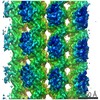
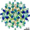
| Title | 4R-MAP4, kinesin-1, and microtubule complex | |||||||||
 Map data Map data | 4R-MAP4%u2013kinesin-1%u2013microtubule complex | |||||||||
 Sample Sample |
| |||||||||
| Biological species |   Bos taurus (cattle) Bos taurus (cattle) | |||||||||
| Method | helical reconstruction / cryo EM / Resolution: 7.35 Å | |||||||||
 Authors Authors | Shigematsu H / Imasaki T / Doki C / Sumi T / Aoki M / Uchikubo-Kamo T / Sakamoto A / Tokuraku K / Shirouzu M / Nitta R | |||||||||
 Citation Citation | Journal: J Cell Biol / Year: 2018 Title: Structural insight into microtubule stabilization and kinesin inhibition by Tau family MAPs. Authors: Hideki Shigematsu / Tsuyoshi Imasaki / Chihiro Doki / Takuya Sumi / Mari Aoki / Tomomi Uchikubo-Kamo / Ayako Sakamoto / Kiyotaka Tokuraku / Mikako Shirouzu / Ryo Nitta /  Abstract: The Tau family microtubule-associated proteins (MAPs) promote microtubule stabilization and regulate microtubule-based motility. They share the C-terminal microtubule-binding domain, which includes ...The Tau family microtubule-associated proteins (MAPs) promote microtubule stabilization and regulate microtubule-based motility. They share the C-terminal microtubule-binding domain, which includes three to five tubulin-binding repeats. Different numbers of repeats formed by alternative splicing have distinct effects on the activities of these proteins, and the distribution of these variants regulates fundamental physiological phenomena in cells. In this study, using cryo-EM, we visualized the MAP4 microtubule complex with the molecular motor kinesin-1. MAP4 bound to the C-terminal domains of tubulins along the protofilaments stabilizes the longitudinal contacts of the microtubule. The strongest bond of MAP4 was found around the intertubulin-dimer interface such that MAP4 coexists on the microtubule with kinesin-1 bound to the intratubulin-dimer interface as well. MAP4, consisting of five repeats, further folds and accumulates above the intertubulin-dimer interface, interfering with kinesin-1 movement. Therefore, these cryo-EM studies reveal new insight into the structural basis of microtubule stabilization and inhibition of kinesin motility by the Tau family MAPs. | |||||||||
| History |
|
- Structure visualization
| Movie |
 Movie viewer Movie viewer |
|---|---|
| Structure viewer | EM map: SurfViewMolmilJmol/JSmol |
| Supplemental images |

- Downloads & links
-EMDB archive
| Map data | emd_9638.map.gz | 13.6 MB | EMDB map data format | |
|---|---|---|---|---|
| Header (meta data) | emd-9638-v30.xmlemd-9638.xml | 10.6 KB 10.6 KB | Display Display | EMDB header |
| Images |  emd_9638.png emd_9638.png | 136.6 KB | ||
| Archive directory |  http://ftp.pdbj.org/pub/emdb/structures/EMD-9638ftp://ftp.pdbj.org/pub/emdb/structures/EMD-9638 http://ftp.pdbj.org/pub/emdb/structures/EMD-9638ftp://ftp.pdbj.org/pub/emdb/structures/EMD-9638 | HTTPS FTP |
-Related structure data


-Links
| EMDB pages | EMDB (EBI/PDBe) / EMDataResource |
|---|
-Map
| File | Download / File: emd_9638.map.gz / Format: CCP4 / Size: 14.7 MB / Type: IMAGE STORED AS FLOATING POINT NUMBER (4 BYTES) | ||||||||||||||||||||||||||||||||||||||||||||||||||||||||||||
|---|---|---|---|---|---|---|---|---|---|---|---|---|---|---|---|---|---|---|---|---|---|---|---|---|---|---|---|---|---|---|---|---|---|---|---|---|---|---|---|---|---|---|---|---|---|---|---|---|---|---|---|---|---|---|---|---|---|---|---|---|---|
| Annotation | 4R-MAP4%u2013kinesin-1%u2013microtubule complex | ||||||||||||||||||||||||||||||||||||||||||||||||||||||||||||
| Voxel size | X=Y=Z: 1.284 Å | ||||||||||||||||||||||||||||||||||||||||||||||||||||||||||||
| Density |
| ||||||||||||||||||||||||||||||||||||||||||||||||||||||||||||
| Symmetry | Space group: 1 | ||||||||||||||||||||||||||||||||||||||||||||||||||||||||||||
| Details | EMDB XML:
CCP4 map header:
| ||||||||||||||||||||||||||||||||||||||||||||||||||||||||||||
-Supplemental data
- Sample components
-Entire : Microtubule-Kinesin1-4R-MAP4 complex
| Entire | Name: Microtubule-Kinesin1-4R-MAP4 complex |
|---|---|
| Components |
|
-Supramolecule #1: Microtubule-Kinesin1-4R-MAP4 complex
| Supramolecule | Name: Microtubule-Kinesin1-4R-MAP4 complex / type: complex / ID: 1 / Parent: 0 |
|---|---|
| Source (natural) | Organism: Bos taurus (cattle) |
| Recombinant expression | Organism:  Escherichia coli (E. coli) / Recombinant strain: BL-21(DE3)pLysS / Recombinant plasmid: pET21d Escherichia coli (E. coli) / Recombinant strain: BL-21(DE3)pLysS / Recombinant plasmid: pET21d |
-Experimental details
-Structure determination
| Method | cryo EM |
|---|---|
 Processing Processing | helical reconstruction |
| Aggregation state | filament |
-Sample preparation
| Buffer | pH: 7.5 / Details: 100mM PIPES-KOH at pH 6.8, 1mM MgCl2, 1mM EGTA |
|---|---|
| Grid | Model: Quantifoil R2/2 / Material: COPPER / Mesh: 300 / Pretreatment - Type: GLOW DISCHARGE |
| Vitrification | Cryogen name: ETHANE / Chamber humidity: 100 % / Chamber temperature: 300 K / Instrument: FEI VITROBOT MARK IV / Details: Blot time 5sec, Blot Force 20. |
- Electron microscopy
| Microscope | FEI TECNAI ARCTICA |
|---|---|
| Electron beam | Acceleration voltage: 200 kV / Electron source: FIELD EMISSION GUN |
| Electron optics | C2 aperture diameter: 30.0 µm / Illumination mode: FLOOD BEAM / Imaging mode: BRIGHT FIELDBright-field microscopy / Nominal defocus max: 2.5 µm / Nominal defocus min: 1.5 µm / Nominal magnification: 78000 |
| Sample stage | Specimen holder model: OTHER / Cooling holder cryogen: NITROGEN |
| Details | Alignment procedure by FEI User Interface Software. Not determined the residual tilt value. |
| Image recording | Film or detector model: FEI FALCON II (4k x 4k) / Detector mode: INTEGRATING / Digitization - Frames/image: 1-34 / Number grids imaged: 1 / Average exposure time: 2.0 sec. / Average electron dose: 55.0 e/Å2 Details: Images were collected in movie-mode at 17 frames per second by using Falcon Hack developed by Greg McMullan |
| Experimental equipment | 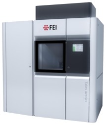 Model: Talos Arctica / Image courtesy: FEI Company |
-Image processing
| CTF correction | Software - Name: CTFFIND (ver. 3) |
|---|---|
| Final angle assignment | Type: NOT APPLICABLE |
| Final reconstruction | Applied symmetry - Helical parameters - Δz: 8.7 Å Applied symmetry - Helical parameters - Δ&Phi: -25.76 ° Applied symmetry - Helical parameters - Axial symmetry: C1 (asymmetric) Resolution.type: BY AUTHOR / Resolution: 7.35 Å / Resolution method: FSC 0.143 CUT-OFF / Number images used: 77616 |
