 Movie
Movie Controller

[English] 日本語
 Yorodumi
- EMDB-9385: The central pair complex of sea urchin (Strongylocentrotus purpur... -
+ Open data

- Basic information
| Entry | Database: EMDB / ID: EMD-9385 | |||||||||
|---|---|---|---|---|---|---|---|---|---|---|
| Title | The central pair complex of sea urchin (Strongylocentrotus purpuratus) sperm resolved by cryo-electron tomography and sub-tomogram averaging | |||||||||
 Map data Map data | The central pair complex of sea urchin (Strongylocentrotus purpuratus) sperm resolved by cryo-electron tomography and sub-tomogram averaging | |||||||||
 Sample Sample |
| |||||||||
| Biological species |   Strongylocentrotus purpuratus (purple sea urchin) Strongylocentrotus purpuratus (purple sea urchin) | |||||||||
| Method | subtomogram averaging / cryo EM / Resolution: 35.0 Å | |||||||||
 Authors Authors | Carbajal-Gonzalez BI / Heuser T / Fu X / Lin J / Smith BW / Mitchell DR / Fu G / Nicastro D | |||||||||
 Citation Citation | Journal: Cytoskeleton (Hoboken) / Year: 2013 Title: Conserved structural motifs in the central pair complex of eukaryotic flagella. Authors: Blanca I Carbajal-González / Thomas Heuser / Xiaofeng Fu / Jianfeng Lin / Brandon W Smith / David R Mitchell / Daniela Nicastro /  Abstract: Cilia and flagella are conserved hair-like appendages of eukaryotic cells that function as sensing and motility generating organelles. Motility is driven by thousands of axonemal dyneins that require ...Cilia and flagella are conserved hair-like appendages of eukaryotic cells that function as sensing and motility generating organelles. Motility is driven by thousands of axonemal dyneins that require precise regulation. One essential motility regulator is the central pair complex (CPC) and many CPC defects cause paralysis of cilia/flagella. Several human diseases, such as immotile cilia syndrome, show CPC abnormalities, but little is known about the detailed three-dimensional (3D) structure and function of the CPC. The CPC is located in the center of typical [9+2] cilia/flagella and is composed of two singlet microtubules (MTs), each with a set of associated projections that extend toward the surrounding nine doublet MTs. Using cryo-electron tomography coupled with subtomogram averaging, we visualized and compared the 3D structures of the CPC in both the green alga Chlamydomonas and the sea urchin Strongylocentrotus at the highest resolution published to date. Despite the evolutionary distance between these species, their CPCs exhibit remarkable structural conservation. We identified several new projections, including those that form the elusive sheath, and show that the bridge has a more complex architecture than previously thought. Organism-specific differences include the presence of MT inner proteins in Chlamydomonas, but not Strongylocentrotus, and different overall outlines of the highly connected projection network, which forms a round-shaped cylinder in algae, but is more oval in sea urchin. These differences could be adaptations to the mechanical requirements of the rotating CPC in Chlamydomonas, compared to the Strongylocentrotus CPC which has a fixed orientation. | |||||||||
| History |
|
- Structure visualization
| Movie |
 Movie viewer Movie viewer |
|---|---|
| Structure viewer | EM map: SurfViewMolmilJmol/JSmol |
| Supplemental images |

- Downloads & links
-EMDB archive
| Map data | emd_9385.map.gz | 3 MB | EMDB map data format | |
|---|---|---|---|---|
| Header (meta data) | emd-9385-v30.xmlemd-9385.xml | 9.9 KB 9.9 KB | Display Display | EMDB header |
| Images |  emd_9385.png emd_9385.png | 45 KB | ||
| Archive directory |  http://ftp.pdbj.org/pub/emdb/structures/EMD-9385ftp://ftp.pdbj.org/pub/emdb/structures/EMD-9385 http://ftp.pdbj.org/pub/emdb/structures/EMD-9385ftp://ftp.pdbj.org/pub/emdb/structures/EMD-9385 | HTTPS FTP |
-Related structure data
| Similar structure data |
|---|


-Links
| EMDB pages | EMDB (EBI/PDBe) / EMDataResource |
|---|
-Map
| File | Download / File: emd_9385.map.gz / Format: CCP4 / Size: 3.8 MB / Type: IMAGE STORED AS FLOATING POINT NUMBER (4 BYTES) | ||||||||||||||||||||||||||||||||||||||||||||||||||||||||||||
|---|---|---|---|---|---|---|---|---|---|---|---|---|---|---|---|---|---|---|---|---|---|---|---|---|---|---|---|---|---|---|---|---|---|---|---|---|---|---|---|---|---|---|---|---|---|---|---|---|---|---|---|---|---|---|---|---|---|---|---|---|---|
| Annotation | The central pair complex of sea urchin (Strongylocentrotus purpuratus) sperm resolved by cryo-electron tomography and sub-tomogram averaging | ||||||||||||||||||||||||||||||||||||||||||||||||||||||||||||
| Voxel size | X=Y=Z: 9.856 Å | ||||||||||||||||||||||||||||||||||||||||||||||||||||||||||||
| Density |
| ||||||||||||||||||||||||||||||||||||||||||||||||||||||||||||
| Symmetry | Space group: 1 | ||||||||||||||||||||||||||||||||||||||||||||||||||||||||||||
| Details | EMDB XML:
CCP4 map header:
| ||||||||||||||||||||||||||||||||||||||||||||||||||||||||||||
-Supplemental data
- Sample components
-Entire : The central pair complex/apparatus (2167 repeats) averaged from 4...
| Entire | Name: The central pair complex/apparatus (2167 repeats) averaged from 41 Strongylocentrotus sea urchin sperms |
|---|---|
| Components |
|
-Supramolecule #1: The central pair complex/apparatus (2167 repeats) averaged from 4...
| Supramolecule | Name: The central pair complex/apparatus (2167 repeats) averaged from 41 Strongylocentrotus sea urchin sperms type: organelle_or_cellular_component / ID: 1 / Parent: 0 |
|---|---|
| Source (natural) | Organism: Strongylocentrotus purpuratus (purple sea urchin) / Organelle: sperm / Location in cell: tail of sperm |
-Experimental details
-Structure determination
| Method | cryo EM |
|---|---|
 Processing Processing | subtomogram averaging |
| Aggregation state | cell |
-Sample preparation
| Buffer | pH: 7.4 / Details: artificial sea water |
|---|---|
| Grid | Model: Quantifoil R2/2 / Material: COPPER / Support film - Material: CARBON / Support film - topology: HOLEY / Pretreatment - Type: GLOW DISCHARGE / Pretreatment - Atmosphere: AIR / Pretreatment - Pressure: 101.325 kPa |
| Vitrification | Cryogen name: ETHANE / Chamber temperature: 298 K / Instrument: HOMEMADE PLUNGER Details: back-side blotting for 1.5-2.5 seconds before plunging. |
| Details | Freshly spawned sea urchin sperms in artificial sea water were used. |
- Electron microscopy
| Microscope | FEI TECNAI F30 |
|---|---|
| Electron beam | Acceleration voltage: 300 kV / Electron source: FIELD EMISSION GUN |
| Electron optics | Calibrated defocus max: 8.0 µm / Calibrated defocus min: 6.0 µm / Calibrated magnification: 13500 / Illumination mode: FLOOD BEAM / Imaging mode: BRIGHT FIELDBright-field microscopy |
| Specialist optics | Energy filter - Name: GIF 2000 / Energy filter - Slit width: 20 eV |
| Sample stage | Specimen holder model: GATAN LIQUID NITROGEN / Cooling holder cryogen: NITROGEN |
| Image recording | Film or detector model: GATAN ULTRASCAN 1000 (2k x 2k) / Average electron dose: 1.5 e/Å2 |
| Experimental equipment | 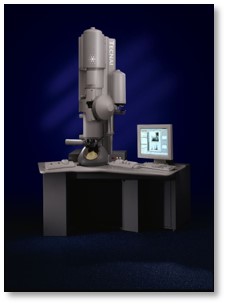 Model: Tecnai F30 / Image courtesy: FEI Company |
-Image processing
| Extraction | Number tomograms: 41 / Number images used: 2167 |
|---|---|
| Final angle assignment | Type: OTHER |
| Final reconstruction | Applied symmetry - Point group: C1 (asymmetric) / Algorithm: BACK PROJECTION / Resolution.type: BY AUTHOR / Resolution: 35.0 Å / Resolution method: FSC 0.5 CUT-OFF / Software - Name: IMOD / Number subtomograms used: 2167 |
