European Molecular Biology Organization (EMBO) Short-term fellowship
ASTF 352-2015
Canadian Institutes of Health Research (CIHR)
Canada
European Molecular Biology Organization (EMBO) Long-term fellowship
ALTF 51-2014
Wellcome Trust
097383
United Kingdom
National Institutes of Health/National Institute of General Medical Sciences (NIH/NIGMS)
PHS GM103314 and PHS GM109824
United States
Biotechnology and Biological Sciences Research Council (BBSRC)
BB/F010656/1
United Kingdom
FEBS long-term fellowship
Robertson Foundation
United States
Fonds de Recherche du Quebec-Sante (FRQ-S)
Canada
Citation
Journal: Nat Commun / Year: 2016 Title: UtpA and UtpB chaperone nascent pre-ribosomal RNA and U3 snoRNA to initiate eukaryotic ribosome assembly. Authors: Mirjam Hunziker / Jonas Barandun / Elisabeth Petfalski / Dongyan Tan / Clémentine Delan-Forino / Kelly R Molloy / Kelly H Kim / Hywel Dunn-Davies / Yi Shi / Malik Chaker-Margot / Brian T ...Authors: Mirjam Hunziker / Jonas Barandun / Elisabeth Petfalski / Dongyan Tan / Clémentine Delan-Forino / Kelly R Molloy / Kelly H Kim / Hywel Dunn-Davies / Yi Shi / Malik Chaker-Margot / Brian T Chait / Thomas Walz / David Tollervey / Sebastian Klinge / Abstract: Early eukaryotic ribosome biogenesis involves large multi-protein complexes, which co-transcriptionally associate with pre-ribosomal RNA to form the small subunit processome. The precise mechanisms ...Early eukaryotic ribosome biogenesis involves large multi-protein complexes, which co-transcriptionally associate with pre-ribosomal RNA to form the small subunit processome. The precise mechanisms by which two of the largest multi-protein complexes-UtpA and UtpB-interact with nascent pre-ribosomal RNA are poorly understood. Here, we combined biochemical and structural biology approaches with ensembles of RNA-protein cross-linking data to elucidate the essential functions of both complexes. We show that UtpA contains a large composite RNA-binding site and captures the 5' end of pre-ribosomal RNA. UtpB forms an extended structure that binds early pre-ribosomal intermediates in close proximity to architectural sites such as an RNA duplex formed by the 5' ETS and U3 snoRNA as well as the 3' boundary of the 18S rRNA. Both complexes therefore act as vital RNA chaperones to initiate eukaryotic ribosome assembly.
History
Deposition
May 27, 2016
-
Header (metadata) release
Jul 6, 2016
-
Map release
Jul 6, 2016
-
Update
Feb 3, 2021
-
Current status
Feb 3, 2021
Processing site: RCSB / Status: Released
-
Structure visualization
Movie
Surface view with section colored by density value
Model: home-made carbon-coated copper grids / Material: COPPER / Support film - Material: CARBON / Support film - topology: CONTINUOUS / Support film - Film thickness: 25.0 nm / Pretreatment - Type: GLOW DISCHARGE
-
Electron microscopy
Microscope
FEI TECNAI 12
Electron beam
Acceleration voltage: 120 kV / Electron source: LAB6
In the structure databanks used in Yorodumi, some data are registered as the other names, "COVID-19 virus" and "2019-nCoV". Here are the details of the virus and the list of structure data.
Jan 31, 2019. EMDB accession codes are about to change! (news from PDBe EMDB page)
EMDB accession codes are about to change! (news from PDBe EMDB page)
The allocation of 4 digits for EMDB accession codes will soon come to an end. Whilst these codes will remain in use, new EMDB accession codes will include an additional digit and will expand incrementally as the available range of codes is exhausted. The current 4-digit format prefixed with “EMD-” (i.e. EMD-XXXX) will advance to a 5-digit format (i.e. EMD-XXXXX), and so on. It is currently estimated that the 4-digit codes will be depleted around Spring 2019, at which point the 5-digit format will come into force.
The EM Navigator/Yorodumi systems omit the EMD- prefix.
Related info.:Q: What is EMD? / ID/Accession-code notation in Yorodumi/EM Navigator
Yorodumi is a browser for structure data from EMDB, PDB, SASBDB, etc.
This page is also the successor to EM Navigator detail page, and also detail information page/front-end page for Omokage search.
The word "yorodu" (or yorozu) is an old Japanese word meaning "ten thousand". "mi" (miru) is to see.
Related info.:EMDB / PDB / SASBDB / Comparison of 3 databanks / Yorodumi Search / Aug 31, 2016. New EM Navigator & Yorodumi / Yorodumi Papers / Jmol/JSmol / Function and homology information / Changes in new EM Navigator and Yorodumi
 Movie
Movie Controller

 Yorodumi
 Open data
 Basic information
 Map data
 Sample
 Saccharomyces cerevisiae (brewer's yeast)
 Authors
 United States,
  Canada,
  United Kingdom, 13 items
  Citation
 Structure visualization
 Movie viewer
 Downloads & links
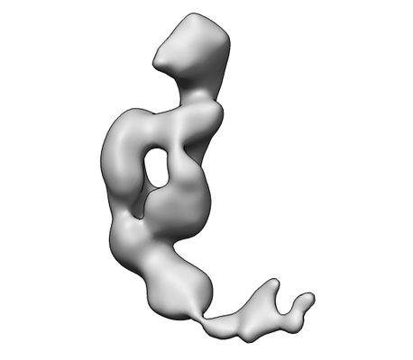 emd_8223.png
 http://ftp.pdbj.org/pub/emdb/structures/EMD-8223


 Sample components
 Processing
 Electron microscopy