 Movie
Movie Controller

[English] 日本語
 Yorodumi
- EMDB-43074: Cryo-EM structure of the Mycobacterium smegmatis 70S ribosome in ... -
+ Open data

- Basic information
| Entry |  | ||||||||||||
|---|---|---|---|---|---|---|---|---|---|---|---|---|---|
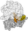
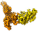
| Title | Cryo-EM structure of the Mycobacterium smegmatis 70S ribosome in complex with hibernation factor Msmeg1130 (Balon) (Structure 4) | ||||||||||||
 Map data Map data | Mycobacterium smegmatis 70S ribosome in complex with hibernation factor Msmeg1130 (Balon). Map | ||||||||||||
 Sample Sample |
| ||||||||||||
 Keywords Keywords |  cryo-EM / mycobacteria / hibernation / Msmeg1130 / Balon / RIBOSOME cryo-EM / mycobacteria / hibernation / Msmeg1130 / Balon / RIBOSOME | ||||||||||||
| Function / homology |  Function and homology information Function and homology informationsmall ribosomal subunit rRNA binding / large ribosomal subunit / regulation of translation / cytosolic small ribosomal subunit / large ribosomal subunit rRNA binding / small ribosomal subunit / 5S rRNA binding / transferase activity / cytosolic large ribosomal subunit / tRNA binding ...small ribosomal subunit rRNA binding / large ribosomal subunit / regulation of translation / cytosolic small ribosomal subunit / large ribosomal subunit rRNA binding / small ribosomal subunit / 5S rRNA binding / transferase activity / cytosolic large ribosomal subunit / tRNA binding / rRNA binding / ribosome / structural constituent of ribosome / ribonucleoprotein complex / translation / mRNA binding / zinc ion binding / metal ion binding / cytoplasmSimilarity search - Function | ||||||||||||
| Biological species |  Mycolicibacterium smegmatis MC2 155 (bacteria) Mycolicibacterium smegmatis MC2 155 (bacteria) | ||||||||||||
| Method | single particle reconstruction / cryo EM / Resolution: 3.1 Å | ||||||||||||
 Authors Authors | Rybak MY / Helena-Bueno K / Hill CH / Melnikov SV / Gagnon MG | ||||||||||||
| Funding support |  United States, 3 items United States, 3 items
| ||||||||||||
 Citation Citation | Journal: Nature / Year: 2024 Title: A new family of bacterial ribosome hibernation factors. Authors: Karla Helena-Bueno / Mariia Yu Rybak / Chinenye L Ekemezie / Rudi Sullivan / Charlotte R Brown / Charlotte Dingwall / Arnaud Baslé / Claudia Schneider / James P R Connolly / James N Blaza / ...Authors: Karla Helena-Bueno / Mariia Yu Rybak / Chinenye L Ekemezie / Rudi Sullivan / Charlotte R Brown / Charlotte Dingwall / Arnaud Baslé / Claudia Schneider / James P R Connolly / James N Blaza / Bálint Csörgő / Patrick J Moynihan / Matthieu G Gagnon / Chris H Hill / Sergey V Melnikov /   Abstract: To conserve energy during starvation and stress, many organisms use hibernation factor proteins to inhibit protein synthesis and protect their ribosomes from damage. In bacteria, two families of ...To conserve energy during starvation and stress, many organisms use hibernation factor proteins to inhibit protein synthesis and protect their ribosomes from damage. In bacteria, two families of hibernation factors have been described, but the low conservation of these proteins and the huge diversity of species, habitats and environmental stressors have confounded their discovery. Here, by combining cryogenic electron microscopy, genetics and biochemistry, we identify Balon, a new hibernation factor in the cold-adapted bacterium Psychrobacter urativorans. We show that Balon is a distant homologue of the archaeo-eukaryotic translation factor aeRF1 and is found in 20% of representative bacteria. During cold shock or stationary phase, Balon occupies the ribosomal A site in both vacant and actively translating ribosomes in complex with EF-Tu, highlighting an unexpected role for EF-Tu in the cellular stress response. Unlike typical A-site substrates, Balon binds to ribosomes in an mRNA-independent manner, initiating a new mode of ribosome hibernation that can commence while ribosomes are still engaged in protein synthesis. Our work suggests that Balon-EF-Tu-regulated ribosome hibernation is a ubiquitous bacterial stress-response mechanism, and we demonstrate that putative Balon homologues in Mycobacteria bind to ribosomes in a similar fashion. This finding calls for a revision of the current model of ribosome hibernation inferred from common model organisms and holds numerous implications for how we understand and study ribosome hibernation. | ||||||||||||
| History |
|
- Structure visualization
| Supplemental images |
|---|

- Downloads & links
-EMDB archive
| Map data | emd_43074.map.gz | 256.7 MB | EMDB map data format | |
|---|---|---|---|---|
| Header (meta data) | emd-43074-v30.xmlemd-43074.xml | 84.6 KB 84.6 KB | Display Display | EMDB header |
| Images | 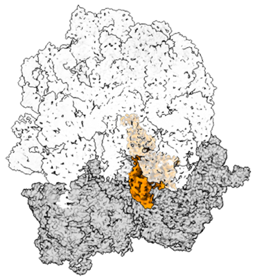 emd_43074.png emd_43074.png | 157.6 KB | ||
| Masks | emd_43074_msk_1.map | 512 MB | Mask map | |
| Filedesc metadata | emd-43074.cif.gz | 15.6 KB | ||
| Others | emd_43074_additional_1.map.gzemd_43074_half_map_1.map.gzemd_43074_half_map_2.map.gz | 459.1 MB 474.7 MB 474.7 MB | ||
| Archive directory |  http://ftp.pdbj.org/pub/emdb/structures/EMD-43074ftp://ftp.pdbj.org/pub/emdb/structures/EMD-43074 http://ftp.pdbj.org/pub/emdb/structures/EMD-43074ftp://ftp.pdbj.org/pub/emdb/structures/EMD-43074 | HTTPS FTP |
-Related structure data
| Related structure data |  8v9jMC  8rd8C  8rdvC  8rdwC  8v9kC  8v9lC M: atomic model generated by this map C: citing same article ( |
|---|---|
| Similar structure data |


-Links
| EMDB pages | EMDB (EBI/PDBe) / EMDataResource |
|---|---|
| Related items in Molecule of the Month |


-Map
| File | Download / File: emd_43074.map.gz / Format: CCP4 / Size: 512 MB / Type: IMAGE STORED AS FLOATING POINT NUMBER (4 BYTES) | ||||||||||||||||||||
|---|---|---|---|---|---|---|---|---|---|---|---|---|---|---|---|---|---|---|---|---|---|
| Annotation | Mycobacterium smegmatis 70S ribosome in complex with hibernation factor Msmeg1130 (Balon). Map | ||||||||||||||||||||
| Voxel size | X=Y=Z: 0.85 Å | ||||||||||||||||||||
| Density |
| ||||||||||||||||||||
| Symmetry | Space group: 1 | ||||||||||||||||||||
| Details | EMDB XML:
|
-Supplemental data
-Mask #1
| File | emd_43074_msk_1.map | ||||||||||||
|---|---|---|---|---|---|---|---|---|---|---|---|---|---|
| Projections & Slices |
| ||||||||||||
| Density Histograms |
 Z
Z Y
Y X
X


-Additional map: Mycobacterium smegmatis 70S ribosome in complex with hibernation...
| File | emd_43074_additional_1.map | ||||||||||||
|---|---|---|---|---|---|---|---|---|---|---|---|---|---|
| Annotation | Mycobacterium smegmatis 70S ribosome in complex with hibernation factor Msmeg1130 (Balon). Sharpened map | ||||||||||||
| Projections & Slices |
| ||||||||||||
| Density Histograms |


-Half map: Mycobacterium smegmatis 70S ribosome in complex with hibernation...
| File | emd_43074_half_map_1.map | ||||||||||||
|---|---|---|---|---|---|---|---|---|---|---|---|---|---|
| Annotation | Mycobacterium smegmatis 70S ribosome in complex with hibernation factor Msmeg1130 (Balon). Half-map 2 | ||||||||||||
| Projections & Slices |
| ||||||||||||
| Density Histograms |


-Half map: Mycobacterium smegmatis 70S ribosome in complex with hibernation...
| File | emd_43074_half_map_2.map | ||||||||||||
|---|---|---|---|---|---|---|---|---|---|---|---|---|---|
| Annotation | Mycobacterium smegmatis 70S ribosome in complex with hibernation factor Msmeg1130 (Balon). Half map 1 | ||||||||||||
| Projections & Slices |
| ||||||||||||
| Density Histograms |


- Sample components
+Entire : Cryo-EM structure of the Mycobacterium smegmatis 70S ribosome in ...
+Supramolecule #1: Cryo-EM structure of the Mycobacterium smegmatis 70S ribosome in ...
+Macromolecule #1: 16S Ribosomal RNA
+Macromolecule #22: poly-U mRNA
+Macromolecule #23: pe/E deacylated phenylanaline-tRNA
+Macromolecule #25: 23S Ribosomal RNA
+Macromolecule #26: 5S Ribosomal RNA
+Macromolecule #2: 30S Ribosomal Protein S2
+Macromolecule #3: 30S ribosomal protein S3
+Macromolecule #4: 30S ribosomal protein S4
+Macromolecule #5: 30S Ribosomal Protein S5
+Macromolecule #6: 30S ribosomal protein S6
+Macromolecule #7: 30S ribosomal protein S7
+Macromolecule #8: 30S Ribosomal Protein S8
+Macromolecule #9: 30S ribosomal protein S9
+Macromolecule #10: 30S ribosomal protein S10
+Macromolecule #11: 30S ribosomal protein S11
+Macromolecule #12: 30S ribosomal protein S12
+Macromolecule #13: 30S ribosomal protein S13
+Macromolecule #14: 30S Ribosomal Protein S14
+Macromolecule #15: 30S Ribosomal Protein S15
+Macromolecule #16: 30S ribosomal protein S16
+Macromolecule #17: 30S ribosomal protein S17
+Macromolecule #18: 30S Ribosomal Protein S18
+Macromolecule #19: 30S ribosomal protein S19
+Macromolecule #20: 30S ribosomal protein S20
+Macromolecule #21: 30S Ribosomal Protein S22
+Macromolecule #24: Ribosome hibernation factor Balon (MSMEG_1130)
+Macromolecule #27: 50S ribosomal protein L2
+Macromolecule #28: 50S ribosomal protein L3
+Macromolecule #29: 50S Ribosomal Protein L4
+Macromolecule #30: 50S Ribosomal Protein L5
+Macromolecule #31: 50S ribosomal protein L6
+Macromolecule #32: 50S ribosomal protein L9
+Macromolecule #33: 50S ribosomal protein L10
+Macromolecule #34: 50S ribosomal protein L11
+Macromolecule #35: 50S Ribosomal Protein L01
+Macromolecule #36: 50S Ribosomal Protein L13
+Macromolecule #37: 50S ribosomal protein L14
+Macromolecule #38: 50S ribosomal protein L15
+Macromolecule #39: 50S Ribosomal Protein L16
+Macromolecule #40: 50S ribosomal protein L17
+Macromolecule #41: 50S Ribosomal Protein L18
+Macromolecule #42: 50S ribosomal protein L19
+Macromolecule #43: 50S Ribosomal Protein L20
+Macromolecule #44: 50S Ribosomal Protein L21
+Macromolecule #45: 50S Ribosomal Protein L22
+Macromolecule #46: 50S Ribosomal Protein L23
+Macromolecule #47: 50S ribosomal protein L24
+Macromolecule #48: 50S ribosomal protein L25
+Macromolecule #49: 50S ribosomal protein L27
+Macromolecule #50: 50S Ribosomal Protein L28
+Macromolecule #51: 50S ribosomal protein L29
+Macromolecule #52: 50S ribosomal protein L30
+Macromolecule #53: 50S Ribosomal Protein L31
+Macromolecule #54: 50S ribosomal protein L32
+Macromolecule #55: 50S Ribosomal Protein L33
+Macromolecule #56: 50S ribosomal protein L34
+Macromolecule #57: 50S ribosomal protein L35
+Macromolecule #58: 50S ribosomal protein L36
+Macromolecule #59: 50S Ribosomal Protein L37
+Macromolecule #60: ZINC ION
-Experimental details
-Structure determination
| Method | cryo EM |
|---|---|
 Processing Processing | single particle reconstruction |
| Aggregation state | particle |
-Sample preparation
| Buffer | pH: 7.5 Details: 20 mM HEPES-KOH, 60 mM KCl, 10 mM MgCl2, 1 mM dithiothreitol |
|---|---|
| Grid | Model: Quantifoil R2/1 / Material: GOLD / Mesh: 200 / Support film - Material: GOLD / Support film - topology: HOLEY / Pretreatment - Type: PLASMA CLEANING / Pretreatment - Time: 30 sec. / Pretreatment - Atmosphere: OTHER |
| Vitrification | Cryogen name: ETHANE / Chamber humidity: 85 % / Chamber temperature: 295 K / Instrument: LEICA EM GP |
- Electron microscopy
| Microscope | TFS KRIOS |
|---|---|
| Electron beam | Acceleration voltage: 300 kV / Electron source: FIELD EMISSION GUN |
| Electron optics | C2 aperture diameter: 100.0 µm / Illumination mode: FLOOD BEAM / Imaging mode: BRIGHT FIELDBright-field microscopy / Cs: 2.7 mm / Nominal defocus max: 2.0 µm / Nominal defocus min: 0.8 µm / Nominal magnification: 105000 |
| Sample stage | Specimen holder model: FEI TITAN KRIOS AUTOGRID HOLDER / Cooling holder cryogen: NITROGEN |
| Image recording | Film or detector model: GATAN K3 BIOQUANTUM (6k x 4k) / Detector mode: COUNTING / Number grids imaged: 1 / Number real images: 11031 / Average electron dose: 40.1 e/Å2 |
| Experimental equipment |  Model: Titan Krios / Image courtesy: FEI Company |
-Image processing
| Startup model | Type of model: NONE |
|---|---|
| Initial angle assignment | Type: MAXIMUM LIKELIHOOD / Software - Name: cryoSPARC (ver. 4.2.1) |
| Final 3D classification | Software - Name: cryoSPARC (ver. 4.2.1) |
| Final angle assignment | Type: MAXIMUM LIKELIHOOD / Software - Name: cryoSPARC (ver. 4.2.1) |
| Final reconstruction | Applied symmetry - Point group: C1 (asymmetric) / Resolution.type: BY AUTHOR / Resolution: 3.1 Å / Resolution method: FSC 0.143 CUT-OFF / Software - Name: cryoSPARC (ver. 4.2.1) / Number images used: 302401 |

