 Movie
Movie Controller

[English] 日本語
 Yorodumi
- EMDB-42855: Cryo-EM structure of the unliganded hexameric prenyltransferase i... -
+ Open data

- Basic information
| Entry |  | |||||||||
|---|---|---|---|---|---|---|---|---|---|---|
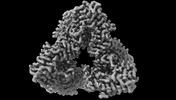
| Title | Cryo-EM structure of the unliganded hexameric prenyltransferase in bifunctional copalyl diphosphate synthase from Penicillium fellutanum with an open conformation, reconstruction in C1 | |||||||||
 Map data Map data | ||||||||||
 Sample Sample |
| |||||||||
 Keywords Keywords |  terpene / biosynthesis / enzyme / TRANSFERASE terpene / biosynthesis / enzyme / TRANSFERASE | |||||||||
| Biological species |  Penicillium fellutanum ATCC 48694 (fungus) Penicillium fellutanum ATCC 48694 (fungus) | |||||||||
| Method | single particle reconstruction / cryo EM / Resolution: 2.94 Å | |||||||||
 Authors Authors | Gaynes MN / Christianson DW | |||||||||
| Funding support |  United States, 1 items United States, 1 items
| |||||||||
 Citation Citation | Journal: J Struct Biol / Year: 2024 Title: Structure of the prenyltransferase in bifunctional copalyl diphosphate synthase from Penicillium fellutanum reveals an open hexamer conformation. Authors: Matthew N Gaynes / Trey A Ronnebaum / Kollin Schultz / Jacque L Faylo / Ronen Marmorstein / David W Christianson / Abstract: Copalyl diphosphate synthase from Penicillium fellutanum (PfCPS) is an assembly-line terpene synthase that contains both prenyltransferase and class II cyclase activities. The prenyltransferase ...Copalyl diphosphate synthase from Penicillium fellutanum (PfCPS) is an assembly-line terpene synthase that contains both prenyltransferase and class II cyclase activities. The prenyltransferase catalyzes processive chain elongation reactions using dimethylallyl diphosphate and three equivalents of isopentenyl diphosphate to yield geranylgeranyl diphosphate, which is then utilized as a substrate by the class II cyclase domain to generate copalyl diphosphate. Here, we report the 2.81 Å-resolution cryo-EM structure of the hexameric prenyltransferase of full-length PfCPS, which is surrounded by randomly splayed-out class II cyclase domains connected by disordered polypeptide linkers. The hexamer can be described as a trimer of dimers; surprisingly, one of the three dimer-dimer interfaces is separated to yield an open hexamer conformation, thus breaking the D3 symmetry typically observed in crystal structures of other prenyltransferase hexamers such as wild-type human GGPP synthase (hGGPPS). Interestingly, however, an open hexamer conformation was previously observed in the crystal structure of D188Y hGGPPS, apparently facilitated by hexamer-hexamer packing in the crystal lattice. The cryo-EM structure of the PfCPS prenyltransferase hexamer is the first to reveal that an open conformation can be achieved even in the absence of a point mutation or interaction with another hexamer. Even though PfCPS octamers are not detected, we suggest that the open hexamer conformation represents an intermediate in the hexamer-octamer equilibrium for those prenyltransferases that do exhibit oligomeric heterogeneity. | |||||||||
| History |
|
- Structure visualization
| Supplemental images |
|---|

- Downloads & links
-EMDB archive
| Map data | emd_42855.map.gz | 643.2 MB |  EMDB map data format EMDB map data format | |
|---|---|---|---|---|
| Header (meta data) | emd-42855-v30.xmlemd-42855.xml | 18.1 KB 18.1 KB | Display Display | EMDB header |
| FSC (resolution estimation) | emd_42855_fsc.xml | 19.2 KB | Display | FSC data file |
| Images |  emd_42855.png emd_42855.png | 51.9 KB | ||
| Filedesc metadata | emd-42855.cif.gz | 5.6 KB | ||
| Others | emd_42855_additional_1.map.gzemd_42855_half_map_1.map.gzemd_42855_half_map_2.map.gz | 361.7 MB 676 MB 676 MB | ||
| Archive directory |  http://ftp.pdbj.org/pub/emdb/structures/EMD-42855ftp://ftp.pdbj.org/pub/emdb/structures/EMD-42855 http://ftp.pdbj.org/pub/emdb/structures/EMD-42855ftp://ftp.pdbj.org/pub/emdb/structures/EMD-42855 | HTTPS FTP |
-Related structure data

-Links
| EMDB pages | EMDB (EBI/PDBe) / EMDataResource |
|---|
-Map
| File | Download / File: emd_42855.map.gz / Format: CCP4 / Size: 729 MB / Type: IMAGE STORED AS FLOATING POINT NUMBER (4 BYTES) | ||||||||||||||||||||
|---|---|---|---|---|---|---|---|---|---|---|---|---|---|---|---|---|---|---|---|---|---|
| Voxel size | X=Y=Z: 0.55 Å | ||||||||||||||||||||
| Density |
| ||||||||||||||||||||
| Symmetry | Space group: 1 | ||||||||||||||||||||
| Details | EMDB XML:
|
-Supplemental data
-Additional map: Unsharpened C1 map of the hexameric PfCPS prenyltransferase...
| File | emd_42855_additional_1.map | ||||||||||||
|---|---|---|---|---|---|---|---|---|---|---|---|---|---|
| Annotation | Unsharpened C1 map of the hexameric PfCPS prenyltransferase in non-uniform refinement | ||||||||||||
| Projections & Slices |
| ||||||||||||
| Density Histograms |
 Z
Z Y
Y X
X


-Half map: #1
| File | emd_42855_half_map_1.map | ||||||||||||
|---|---|---|---|---|---|---|---|---|---|---|---|---|---|
| Projections & Slices |
| ||||||||||||
| Density Histograms |


-Half map: #2
| File | emd_42855_half_map_2.map | ||||||||||||
|---|---|---|---|---|---|---|---|---|---|---|---|---|---|
| Projections & Slices |
| ||||||||||||
| Density Histograms |


- Sample components
-Entire : Hexameric prenyltransferase from the bifunctional copalyl diphosp...
| Entire | Name: Hexameric prenyltransferase from the bifunctional copalyl diphosphate synthase of Penicillium fellutanum |
|---|---|
| Components |
|
-Supramolecule #1: Hexameric prenyltransferase from the bifunctional copalyl diphosp...
| Supramolecule | Name: Hexameric prenyltransferase from the bifunctional copalyl diphosphate synthase of Penicillium fellutanum type: complex / ID: 1 / Parent: 0 / Macromolecule list: all |
|---|---|
| Source (natural) | Organism: Penicillium fellutanum ATCC 48694 (fungus) |
| Molecular weight | Theoretical: 634.002 KDa |
-Macromolecule #1: Copalyl diphosphate synthase
| Macromolecule | Name: Copalyl diphosphate synthase / type: protein_or_peptide / ID: 1 / Enantiomer: LEVO |
|---|---|
| Source (natural) | Organism: Penicillium fellutanum ATCC 48694 (fungus) |
| Recombinant expression | Organism:  Escherichia coli BL21(DE3) (bacteria) Escherichia coli BL21(DE3) (bacteria) |
| Sequence | String: MGSSHHHHHH SSGENLYFQG HMASMESASL DQSAALLVKE LTEHIDDSNG LGFMSPAIYD TAWVSMIKKT DNDQTFWLFP KSFHYILENQ LENGGWVTYA SEIDGILNTS ASLLSLKRHF DLPLQISTES QTSMENRIRK ATDALRVLLR TWDVDATLHV GFEILVPALL ...String: MGSSHHHHHH SSGENLYFQG HMASMESASL DQSAALLVKE LTEHIDDSNG LGFMSPAIYD TAWVSMIKKT DNDQTFWLFP KSFHYILENQ LENGGWVTYA SEIDGILNTS ASLLSLKRHF DLPLQISTES QTSMENRIRK ATDALRVLLR TWDVDATLHV GFEILVPALL DYLQVEGLTF DFPGRDKLFQ IRDQKLSRFK PEFIYAPFQT TALHSLEAFI GLIDFDRVQH HKVRGSFMAS PSSTAAVLMN ATEWDIDCEE YIRHVIEHAS GKASGGVPSA FPSTIFEITW TLSTLLKAGF NLSSNDSSNV QKACSYLLGV LTAEKGAIGF VPSVCADADD TAKTILVLSL LRENVLPDGM LKAFEVENHF KTYPLERDPS FSANCNVLLA LLHLENPSQY TLQIEKATRF LYTHFRESNL NVRDKWNLSP FYSWMLMAQA IARLDELCKG SQLKNLHDHV ESDLIPLLQE MTVSVMHQQN KDGSWGTKLS KEETAYAVLM LTYAVSFEAL VGPRRQIRNA IEEGCLFLRS GKDATSERLW VEKVTYESQM LSRAYTLAAL KNALDVLKKD DMKIAFIGNS VNESFTEIEV VNGKNVTEPS SNTYDLKEVK MDKAEFTPPD TPPRSKSTSV DPIEEAICAH ENRGAISDHT SRAIDLCRNP PLWGTDQEQT LLGPFEYLES IPGKNIRSQF IEAFNTWLQI PQDHLQIVGK VISMLHTASL LVDDIEDNSL LRRGQPVAHS IFGTAQTFNS GNYVYFLALQ EVQKLNSPRA ISIFVDALTQ LHRGQGMDVF WRDSLICPTE EEYLDMVANK TGALFCLAIE LLQIKSTVQL DFLPLVRLLG IIFQICDDYL NLKSTNYTQK KGLCEDITEG KFSFPIIHSI RTKPGNRQLI NVLRQKSKED DVKRFALAYM ESTQSFDYTR DFVKILNGEA LRMIEDLEQQ GLHRNIEIRN ILARMSLEQ |
-Experimental details
-Structure determination
| Method | cryo EM |
|---|---|
 Processing Processing | single particle reconstruction |
| Aggregation state | particle |
-Sample preparation
| Concentration | 0.5 mg/mL | ||||||||||||
|---|---|---|---|---|---|---|---|---|---|---|---|---|---|
| Buffer | pH: 7.5 Component:
| ||||||||||||
| Grid | Model: Quantifoil R1.2/1.3 / Material: COPPER / Mesh: 300 | ||||||||||||
| Vitrification | Cryogen name: ETHANE / Chamber humidity: 100 % / Chamber temperature: 277 K / Instrument: FEI VITROBOT MARK I |
- Electron microscopy
| Microscope | FEI TITAN KRIOS |
|---|---|
| Electron beam | Acceleration voltage: 300 kV / Electron source: FIELD EMISSION GUN |
| Electron optics | C2 aperture diameter: 100.0 µm / Illumination mode: FLOOD BEAM / Imaging mode: BRIGHT FIELDBright-field microscopy / Cs: 2.7 mm / Nominal defocus max: 2.5 µm / Nominal defocus min: 0.8 µm |
| Image recording | Film or detector model: GATAN K3 BIOQUANTUM (6k x 4k) / Number real images: 12467 / Average exposure time: 2.15 sec. / Average electron dose: 43.0 e/Å2 |
| Experimental equipment |  Model: Titan Krios / Image courtesy: FEI Company |
-Image processing
| Particle selection | Number selected: 3639075 |
|---|---|
| Startup model | Type of model: INSILICO MODEL |
| Initial angle assignment | Type: MAXIMUM LIKELIHOOD |
| Final angle assignment | Type: MAXIMUM LIKELIHOOD |
| Final reconstruction | Applied symmetry - Point group: C1 (asymmetric) / Resolution.type: BY AUTHOR / Resolution: 2.94 Å / Resolution method: FSC 0.143 CUT-OFF / Number images used: 267283 |
| FSC plot (resolution estimation) |  |
