National Institutes of Health/National Institute of General Medical Sciences (NIH/NIGMS)
GM122510
United States
Citation
Journal: Proc Natl Acad Sci U S A / Year: 2023 Title: Tad and toxin-coregulated pilus structures reveal unexpected diversity in bacterial type IV pili. Authors: Ravi R Sonani / Juan Carlos Sanchez / Joseph K Baumgardt / Shivani Kundra / Elizabeth R Wright / Lisa Craig / Edward H Egelman / Abstract: Type IV pili (T4P) are ubiquitous in both bacteria and archaea. They are polymers of the major pilin protein, which has an extended and protruding N-terminal helix, α1, and a globular C-terminal ...Type IV pili (T4P) are ubiquitous in both bacteria and archaea. They are polymers of the major pilin protein, which has an extended and protruding N-terminal helix, α1, and a globular C-terminal domain. Cryo-EM structures have revealed key differences between the bacterial and archaeal T4P in their C-terminal domain structure and in the packing and continuity of α1. This segment forms a continuous α-helix in archaeal T4P but is partially melted in all published bacterial T4P structures due to a conserved helix breaking proline at position 22. The tad (tight adhesion) T4P are found in both bacteria and archaea and are thought to have been acquired by bacteria through horizontal transfer from archaea. Tad pilins are unique among the T4 pilins, being only 40 to 60 residues in length and entirely lacking a C-terminal domain. They also lack the Pro22 found in all high-resolution bacterial T4P structures. We show using cryo-EM that the bacterial tad pilus from is composed of continuous helical subunits that, like the archaeal pilins, lack the melted portion seen in other bacterial T4P and share the packing arrangement of the archaeal T4P. We further show that a bacterial T4P, the toxin coregulated pilus, which lacks Pro22 but is not in the tad family, has a continuous N-terminal α-helix, yet its α1 s are arranged similar to those in other bacterial T4P. Our results highlight the role of Pro22 in helix melting and support an evolutionary relationship between tad and archaeal T4P.
In the structure databanks used in Yorodumi, some data are registered as the other names, "COVID-19 virus" and "2019-nCoV". Here are the details of the virus and the list of structure data.
Jan 31, 2019. EMDB accession codes are about to change! (news from PDBe EMDB page)
EMDB accession codes are about to change! (news from PDBe EMDB page)
The allocation of 4 digits for EMDB accession codes will soon come to an end. Whilst these codes will remain in use, new EMDB accession codes will include an additional digit and will expand incrementally as the available range of codes is exhausted. The current 4-digit format prefixed with “EMD-” (i.e. EMD-XXXX) will advance to a 5-digit format (i.e. EMD-XXXXX), and so on. It is currently estimated that the 4-digit codes will be depleted around Spring 2019, at which point the 5-digit format will come into force.
The EM Navigator/Yorodumi systems omit the EMD- prefix.
Related info.:Q: What is EMD? / ID/Accession-code notation in Yorodumi/EM Navigator
Yorodumi is a browser for structure data from EMDB, PDB, SASBDB, etc.
This page is also the successor to EM Navigator detail page, and also detail information page/front-end page for Omokage search.
The word "yorodu" (or yorozu) is an old Japanese word meaning "ten thousand". "mi" (miru) is to see.
Related info.:EMDB / PDB / SASBDB / Comparison of 3 databanks / Yorodumi Search / Aug 31, 2016. New EM Navigator & Yorodumi / Yorodumi Papers / Jmol/JSmol / Function and homology information / Changes in new EM Navigator and Yorodumi
 Movie
Movie Controller

 Yorodumi
 Open data
 Basic information
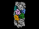
 Map data
 Sample
 Keywords
 Toxin /
  Function and homology information

 Authors
 United States, 1 items
  Citation
 Structure visualization
 Downloads & links
 emd_42279.png
 http://ftp.pdbj.org/pub/emdb/structures/EMD-42279


 Z
Z Y
Y X
X


 Sample components
 Processing
 Electron microscopy

